 |
|
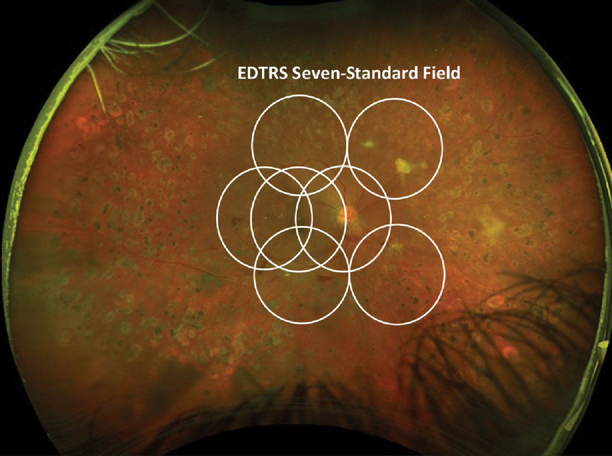
Despite finding similarities in disease worsening regardless of the distribution of lesions, researchers noted that more extensive lesions identified beyond the ETDRS seven-standard fields were associated with a greater risk of disease worsening. Photo: Julie Torbit, OD, and Brad Sutton, OD. Click image to enlarge. |
The Early Treatment Diabetic Retinopathy Study (EDTRS) was a watershed moment in retinal disease care and its results have informed the management of diabetic eye disease ever since. However, it was performed over 30 years ago and thus fails to account for a host of more recent research and newer technologies. The EDTRS Diabetic Retinopathy Severity Scale (DRSS) is still widely used but its seven-standard field imaging basis may miss evidence of DR in the periphery. Today, physicians can use ultra-widefield (UWF) photography or fluorescein angiography to better determine the risk of worsening DRSS scores based on the type and distribution of the disease.
A study presented at ARVO 2024 in Seattle yesterday demonstrated how researchers evaluated the effect of diabetic retinopathy lesions using these newer modalities and the results they observed. In a multicenter prospective longitudinal observational study, researchers examined 544 eyes with nonproliferative diabetic retinopathy and DRSS scores between levels 35 and 53. The primary outcome for the study was found by using UWF color and fluorescein angiography imaging to determine disease worsening, which is defined by either DRSS worsening by ≥2 steps from baseline after imaging or the eventual involvement of diabetic retinopathy treatment over a four year period.
Researchers examined their patients over a four-year period while observing specific DR lesions. Types of lesions investigated for the study included hemorrhages and/or microaneurysms, intraretinal microvascular abnormalities, new vessels elsewhere and venous beading.
After four years, the researchers found that disease worsening was similar in all cases despite where the hemorrhages and/or microaneurysms or intraretinal microvascular abnormalities were located in the retina. More severe lesion grades were found for hemorrhages and/or microaneurysms when assessed using UWF color imaging . When imaged with UWF technologies, researchers noted greater risks of disease progression at the following hazard ratios (HR) based on lesion type:
- hemorrhages and/or microaneurysms, HR = 1.74 on UWF photography and HR = 1.90 on UWF angiography
- intraretinal microvascular abnormalities, HR = 1.68 on UWF angiography
- new vessels elsewhere, HR 1.99 on UWF angiography
Venous beading, however, was not greatly associated with disease worsening, according to the ARVO abstract.
The researchers characterized the importance of updating one’s clinical protocols for screening and following DR patients. “This finding suggests that failing to identify eyes that have peripheral diabetic retinopathy lesions may lead to underestimation of risk of diabetic retinopathy worsening and that identification of lesions throughout the entire retina is important to more accurately stratify risk for progression over time,” mentioned the researchers in their ARVO abstract.
Original abstract content ©2024 Association for Research in Vision and Ophthalmology.
Silva PS. Diabetic retinopathy lesion types and distribution on ultrawide field imaging and the risk of disease worsening over time. ARVO 2024 annual meeting. |