Innovations in Eye CareCheck out the other feature articles in this month's issue:A Light in the DARC: Seeing Glaucoma Before it Strikes Bioengineering the Retinal Pigment Epithelium Iontophoresis: Wave of the Future? While You Were Sleeping |
As eye care professionals, we diagnose patients with primary open angle glaucoma (POAG) every day in our clinics. We tell them about the disease and how the pattern of vision loss begins with the peripheral vision and slowly encroaches centrally. We tell them they will need to begin lifelong treatment with an antihypertensive drop and possibly laser therapy. The indolent progression of glaucoma often leaves patients understandably surprised by their diagnosis, as the vision loss they have experienced is hidden to them.
Patients commonly ask if there are other treatment options for their condition. We impress on our patients that drops are currently the first-line treatment for glaucoma. After their initial pressure checks, patients return to us at their six-month and one-year follow-ups describing difficulties with their drops. “I tried the drops but I didn’t like them,” “I forgot to take them,” and “they make my eyes uncomfortable” are all common explanations for non-adherence. These reasons are always troubling because for many patients, conventional treatments seem unacceptable or impractical to incorporate into their daily lives.
Over the past 15 years, glaucoma management has begun to shift. Clinicians are moving away from treating patients with topical drops to maximum medical therapy then referring for traditional filtering surgery, which is traumatic and carries significant risk. Instead, we have seen the rise of minimally invasive glaucoma surgeries (MIGS), which are less invasive and possess greater safety profiles than traditional glaucoma surgery.1 Additional benefits include decreasing patients’ medication burden while maintaining intraocular pressure (IOP) control. However, little progress has been made for patients diagnosed with milder stages of disease.
The IOPTx system from Bionode is aiming to offer these patients a solution in the form of a contact lens and glasses combination designed to lower intraocular pressure. Our clinicians at the Prism Eye Institute are evaluating these efforts.
How It Works
Previous research has shown transcorneal stimulation is safe for the anterior segment and deeper structures of the globe in the treatment of retinitis pigmentosa and various optic neuropathies.2-5 Bionode has used this research as the underlying basis in developing IOPTx, which uses transcorneal electrical stimulation that targets the aqueous inflow and outflow structures of the eye in an effort to reduce IOP.
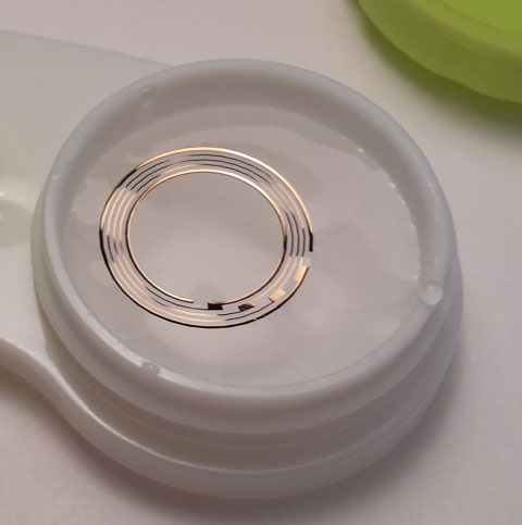 |
| This gold coil, affixed to a contact lens, is triggered by an electromagnetic field and delivers a stimulus that, researchers speculate, could reduce IOP. Photo: Bionode |
The system consists of a lightweight pair of glasses and customized contact lenses. The spectacles have embedded electronics in addition to a circuit that delivers an electromagnetic stimulus that reduces IOP. These glasses are fitted with wound enamel copper wire–coated coils that receive electricity from an external pulse generator and battery pack attached to the coils via a USB cable. When the device is activated, the pulse generator in the spectacle coil creates an electromagnetic field directed towards the eye and induces a current in the customized contact lens.
The contact lens consists of two Alcon Air Optix hydrophilic hydrogel contact lenses (Alcon) with the gold coil embedded in an electrically insulating parylene substrate between the two lenses. The contact lens acts as a secondary coil that is stimulated by the electromagnetic field emitted from the spectacle coil inserts. The electricity generated is driven across the physiologic structures of the eye.
Another version of the IOPTx system bypasses the use of a contact lens altogether and instead uses the electromagnetic field generated by a coil on the glasses as the only neuromodulatory source to potentially lower IOP. This may be useful in patients who cannot tolerate contact lenses, although currently this version of the device is not included in any clinical trials.
Based on animal and biomechanical studies, the purported mechanism of action is twofold. First, the primary IOP-reducing effect stems from the electrical current causing interference with the normal functioning of the ciliary epithelial ion pumps. This, in turn, decreases the quantity of aqueous humor actively transported into the posterior chamber. The secondary mechanism of action involves electrical stimulation of the ciliary body causing contraction of the muscle and opening of the drainage structures of Schelmm’s Canal. Potentially, this means the device is able to modulate both the outflow and production of aqueous simultaneously.
Multicenter Clinical Trials
Clinical trials of the device are currently underway in Spain and are beginning at the Prism Eye Institute in Mississauga, Canada. The study design is a prospective multicenter double-masked randomized clinical trial. At the Mississauga site, we are hoping to enroll 20 to 30 patients with POAG or ocular hypertension. After a washout period, patients will be randomized to the Bionode and control groups. Exclusion criteria will include secondary types of glaucoma and patients with previous glaucoma surgery. Enrolled patients will undergo treatment using the Bionode IOPTx system and IOP will be measured periodically over one month. The primary objective of the study is to evaluate the safety and IOP-lowering efficacy of the IOPTx system in glaucomatous eyes. Secondary outcomes will include the longevity of IOP reduction. This trial will provide valuable information for the appropriate use and protocol for the device in patients with POAG.
Difficulties with Traditional Glaucoma Treatment
Non-adherence is a long-standing problem in glaucoma management.6 Recent research shows that between 1995 and 2001 in the United States almost half of patients who filled one prescription discontinued their use after the first bottle and only 37% continued use at three years.7 Another study—one that used electronic monitoring systems—published in 2015 compared patients’ self-reported medication adherence to medication event monitors at 60-day follow-up appointments. In this study, 31% overestimated their drop use when compared with their monitors.8 In the future, the IOPTx system could be used to address patient compliance issues and, hopefully, reduce glaucoma progression.
Ocular surface disease (OSD) is one of the most frequent comorbidities associated with glaucoma, as nearly 50% of patients treated with topical glaucoma medications have OSD.9 Topical medications often contain preservatives, such as benzaklkonium chloride, which is cytotoxic and can destroy normal conjunctival epithelial cell membrane, ultimately releasing proinflammatory mediators that can lead to inflammation and dryness and be a precipitating factor for OSD.10 Continual use of topical medications can lead to alterations in tear film composition and even damage to the ocular surface.10
Preservative-free medications are available to combat this problem; however, these medications are not as readily available and increase the cost to patients. They also do not address patient compliance issues. Neuromodulation may have a role in reducing rates of ocular surface disease related to topical hypotensive medication in glaucoma patients.
Another option is laser trabeculoplasty. This is an appropriate, noninvasive option as a primary or secondary treatment for POAG or in patients who have adherence issues with medication. Selective laser trabeculoplasty (SLT) can achieve 20% or greater reduction in IOP in 58% to 94% of patients at 12 months.11 However, the attrition rate is high, with only 38% to 68% of eyes maintaining 20% reduction at four years.11
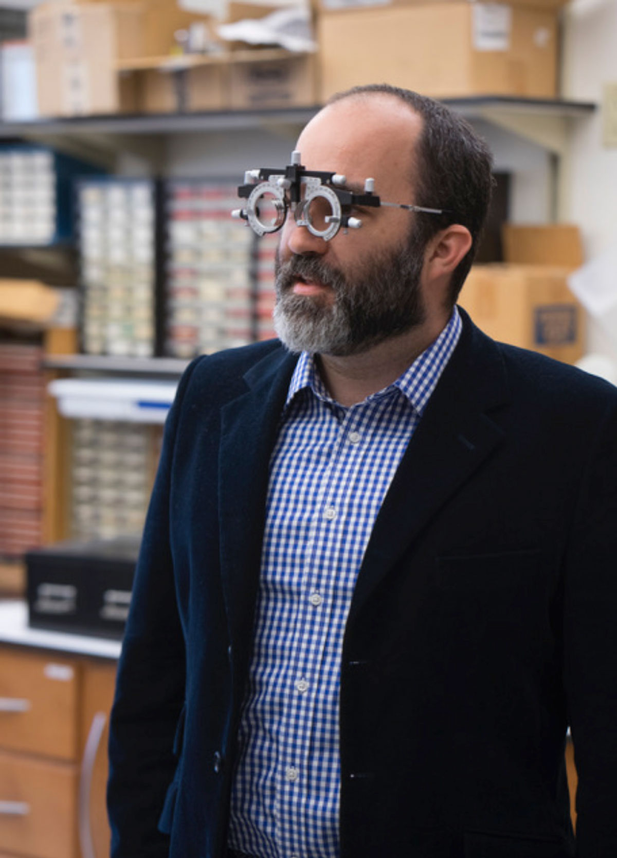 |
| The Bionode system uses spectacles embedded with circuitry to deliver an electromagnetic stimulus designed to reduce IOP. Photo: Bionode |
Wearables
Recent trends in medicine have sought to develop treatment and monitoring modalities personalized to patients’ needs. Some of the emerging wearable noninvasive devices aim to monitor and treat patients with glaucoma.
Triggerfish contact lens sensor (CLS) (Sensimed). This is a noninvasive wireless soft contact lens that measures IOP over a 24-hour period. The CLS sends IOP measurements wirelessly to an adhesive disposable antenna that is placed around the periorbital skin. The antenna is connected to a recorder the patient wears in a pouch, similar to 24-hour blood pressure monitor. The CLS measures changes in corneal curvature to estimate IOP over a 24-hour period. This device has the potential to capture the variation of a patient’s IOP during their daily activities, as opposed to merely when they are in the office. This could help identify their true peak IOP, which is associated with long-term progression of their disease.12 A recent study of this technology concluded that the CLS appears to be better than mean clinic Goldmann IOP measurements at assessing risk of glaucomatous visual field deterioration.13
Repetitive transorbital alternating current stimulation (rtACS). Alternating current stimulation of the brain has been shown to potentially increase excitability and synchronicity in patients with brain injury.14-16 This method of neuromodulation has recently been applied to the visual system in the context of optic neuropathy. A small study involving patients with non-specific optic neuropathies showed that rtACS was able to improve patients’ visual fields in the treatment arm.17 This study was limited by the small number of enrolled patients (n=12) in the treatment group, but its application in glaucoma management is promising. A larger prospective randomized double blind, sham-controlled trial compared rtACS to placebo in partially blind patients with glaucoma (n=33) or other causes of optic nerve damage (n=50) and demonstrated a 24% improvement in visual fields in the treatment group.18 In the future, this technology could possibly maximize the residual visual potential of patients with glaucoma who have significant loss of visual field.
IOP modulating goggles. Researchers suggest significant interplay between the pressure of the cerebrospinal fluid (CSF) and the optic nerve.19,20 A correlation also exists between low CSF pressure and the development of glaucoma.19,20 Although the theoretical basis behind the pathogenesis of glaucoma in this patient population is still under debate, supporters postulate that patients with low CSF pressure have an abnormally high trans–lamina cribrosa pressure difference that leads to glaucomatous nerve damage.20
Balance Goggles (Equinox) attempt to balance the pressure differential by applying a negative vacuum around the orbit of the eye. The company has received funding from NASA’s National Space Biomedical Research institute and are currently conducting research on up to 50 clinically normal eyes to evaluate safety and efficacy of their product. This technology may have future applications in glaucoma management as well as idiopathic intracranial hypertension, hypotony and visual impairment and intraocular pressure.
Innovation is never easy, but it is necessary to drive disease management forward and overcome barriers to conventional treatment. New technology is moving towards less invasive and more objective measures for chronic diseases such as glaucoma. While further research is needed, the Bionode IOPTx system has the potential to play a promising role in the glaucoma management algorithm.
Drs. Lukasik is a research fellow at the Prism Eye Institute.
Dr. Ahmed practices at the Prism Eye Institute, Mississauga, Canada and is a professor of ophthalmology at the University of Toronto, Department of Ophthalmology and Vision Sciences, Toronto, Canada.
|
1. Pillunat LE, Erb C, Jünemann AG, Kimmich F. Micro-invasive glaucoma surgery (MIGS): a review of surgical procedures using stents. Clin Ophthalmol. 2017;11:1583-600. 2. Naycheva L, Schatz A, Röck T, et al. Phosphene thresholds elicited by transcorneal electrical stimulation in healthy subjects and patients with retinal diseases. Invest Ophthalmol Vis Sci. 2012;53(12):7440-8. 3. Schatz A, Pach J, Gosheva M, et al. Transcorneal electrical stimulation for patients with retinitis pigmentosa: A prospective, randomized, sham-controlled follow-up study over 1 year. Invest Ophthalmol Vis Sci. 2017;58(1):257–69. 4. Fujikado T, Morimoto T, Kanda H, et al. Evaluation of phosphenes elicited by extraocular stimulation in normals and by suprachoroidal-transretinal stimulation in patients with retinitis pigmentosa. Graefes Arch Clin Exp Ophthalmol. 2007;245(10):1411-9. 5. Fujikado T, Morimoto T, Matsushita K, et al. Effect of transcorneal electrical stimulation in patients with nonarteritic ischemic optic neuropathy or traumatic optic neuropathy. Jpn J Ophthalmol. 2006;50(3):266-73. 6. Bloch S, Rosenthal AR, Friedman L, Caldarolla P. Patient compliance in glaucoma. Br J Ophthalmol. 1977;61(8):531-4. 7. Nordstrom BL, Friedman DS, et al. Persistence and adherence with topical glaucoma therapy. Am J Ophthalmol. 2005;140(4):598-606. 8. Sayner R, Carpenter DM, Blalock SJ, et al. Accuracy of patient-reported adherence to glaucoma medications on a visual analog scale compared with electronic monitors. Clinical Therapeutics. 2015;37(9):1975-85. 9. Fechtner RD, Godfrey DG, Budenz D, et al. Prevalence of ocular surface complaints in patients with glaucoma using topical intraocular pressure-lowering medications. Cornea. 2010;29(6):618-21. 10. Mastropasqua L, Agnifili L, Mastropasqua R, Fasanella V. Conjunctival modifications induced by medical and surgical therapies in patients with glaucoma. Curr Opin Pharmacol. 2013;13(1):56-64. 11. Leahy KE, White AJ. Selective laser trabeculoplasty: current perspectives. Clin Ophthalmol. 2015;9:833-41. 12. Konstas AGP, Quaranta L, Mikropoulos DG, et al. Peak intraocular pressure and glaucomatous progression in primary open-angle glaucoma. J Ocul Pharmacol Ther. 2012;28(1):26-32. 13. De Moraes CG, Mansouri K, Liebmann JM, Ritch R, Triggerfish Consortium. Association between 24-Hour intraocular pressure monitored with contact lens sensor and visual field progression in older adults with glaucoma. JAMA Ophthalmol. 2018;136(7):779-85. 14. Nair DG, Renga V, Lindenberg R, et al. Optimizing recovery potential through simultaneous occupational therapy and non-invasive brain-stimulation using tDCS. Restor Neurol Neurosci. 2011;29(6):411-20. 15. Song S, Sandrini M, Cohen LG. Modifying somatosensory processing with non-invasive brain stimulation. Restor Neurol Neurosci. 2011;29(6):427-37. 16. Chrysikou EG, Hamilton RH. Noninvasive brain stimulation in the treatment of aphasia: exploring interhemispheric relationships and their implications for neurorehabilitation. Restor Neurol Neurosci. 2011;29(6):375-94. 17. Sabel BA, Fedorov AB, Naue N, et al. Non-invasive alternating current stimulation improves vision in optic neuropathy. Restor Neurol Neurosci. 2011;29(6):493-505. 18. Gall C, Schmidt S, Schittkowski MP, et al. Alternating current stimulation for vision restoration after optic nerve damage: A randomized clinical trial. PLoS ONE. 2016;11(6):e0156134. 19. Berdahl JP, Fautsch MP, Stinnett SS, Allingham RR. Intracranial pressure in primary open angle glaucoma, normal tension glaucoma, and ocular hypertension: a case-control study. Invest Ophthalmol Vis Sci. 2008;49(12):5412-8. 20. Ren R, Jonas JB, Tian G, et al. Cerebrospinal fluid pressure in glaucoma: a prospective study. Ophthalmology. 2010;117(2):259-66. |
