Cataract surgery is performed about three million times each year in the US, achieving fabulous results for the majority of patients. Unfortunately, approximately 30% to 50% of patients will develop a secondary cataract—also known as posterior capsular opacification (PCO)—at some point following cataract surgery. PCO can develop a few days to several years following cataract surgery, but occurs most commonly within the first one to two years following cataract extraction.
|
|


Patients often describe their vision as “not being as good as it was right after cataract surgery.” Pay special attention to younger patients and those with silicone IOLs, as both of these factors increase the risk of PCO formation.
Qualification criteria for YAG laser capsulotomy closely follows those for cataract surgery itself: most patients should have a best-corrected visual acuity of 20/40 or worse via refraction or glare testing. We often like to document a one- to two-line improvement in vision on potential acuity testing.
At the preoperative exam, it is critical to dilate your patient to give the best view of the PCO, as well as allow for a full dilated retinal exam. Take care to ensure that the macula and peripheral retina are healthy, given the nature of where and how the laser works. If you observe active macular edema, vitreomacular traction or peripheral holes, the procedure is contraindicated until a retinal specialist has cleared the patient.
|
|
|
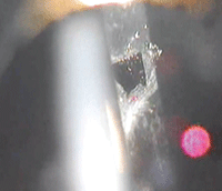
Breakthrough of a Grade 3 PCO by YAG capsulotomy. Note the laser spot on the capsule (small red dot) and the corresponding reflection on the cornea. To watch a narrated video of YAG capsulotomy,
click here.
|
Surgical Protocol
One drop of brimonidine 0.1% is instilled preoperatively to control the risk of IOP spike. A laser lens may be placed on the eye, depending on the laser being used and doctor preference. Proper, continuous patient education is critical during the procedure, as the patient may experience “popping” or an acoustic sound in their ears. This is a normal phenomenon that is experienced with the YAG laser during both peripheral iridotomies and capsulotomies.
Small pulses of YAG laser energy are placed just posterior to the PCO in the anterior vitreous. The energy then travels anteriorly, towards the front of the eye, disrupting the PCO and causing the cloudy membrane to open. Pulses of 1.5mJ to 3.0mJ energy in a single-shot distribution are commonly used to disrupt the PCO, typically in a cruciate or cross-like pattern.
Another drop of brimonidine 0.1% is instilled immediately post-laser to minimize the risk of IOP elevation. The patient is sent home with an anti-inflammatory, often prednisolone acetate 1.0%, to be used QID for one week.
At the one-week post-op visit, a close, undilated look at the IOL and posterior capsule is taken to ensure that no flaps or pieces of capsule remain along the visual axis or pupil, as that can contribute to continued glare or decreased vision. If needed, a touch-up procedure may be performed with the laser at this visit. The patient is also dilated at the one-week post-op exam to rule out any posterior segment problems.
YAG laser capsulotomy is the only effective treatment for posterior capsular opacification. Proper recognition and treatment will almost certainly improve your patient’s vision, and leave them smiling and telling their friends how you returned it to the outstanding level they enjoyed immediately after cataract surgery.
Dr. Lighthizer is an assistant professor and the chief of the specialty care and electrodiagnostics clinics at Oklahoma College of Optometry.
