During a pediatric eye exam, you’ll often discover that the child truly cannot read the chart––even with a parent sitting there saying, “Come on, you can see that!” As you carefully address the situation and perform your refraction, you determine that the child is significantly myopic. Suddenly, you hear the child read “T, Z, V, E, C, L.” Then, when you remove the phoropter, you hear him mutter the word “Wow.” Now, what do you do?
First, attempt to alleviate the guilt and embarrassment that the parent is about to feel. Next, take some time to describe all the options in refractive care. While doing so, educate both the parent and child about the condition of myopia, how it progresses with increased age and how it can impact the child’s performance in both school and sports. Then, be sure to review all the corrective options––from spectacles and conventional contact lenses to pharmaceutical treatments and orthokeratology (ortho-k) lenses (see “ Options for Myopia Control."). Remember to stress the potential visual benefits and financial costs associated with each corrective method. Together, you can help create the best plan to improve the child’s vision.
This article chiefly will focus on the use of ortho-k lenses as a primary treatment for myopia control, an option sometimes overlooked in favor of conventional corrective lenses.
Where to Start?
First, you must reassure the child that he or she is not going blind, but instead will simply require corrective eyewear to achieve the best possible visual function and performance. Additionally, briefly summarize some of the evidence-based medicine associated with the control of myopia progression.
Confirmatory testing is critical prior to detailed planning and discussion. This should include a battery of examinations, such as cycloplegic evaluation, accommodative response and lag, phoria, accommodative convergence/accommodation ratio, intraocular pressure, corneal topography and possibly wavefront aberrometry.1,2 Also, you may wish to screen for predictive signs of juvenile myopia, including cycloplegic refraction, spherical refractive error, axial length and corneal power.3
Early Myopia Development and Progression
In 1989, researchers from the Orinda Longitudinal Study of Myopia (OLSM) analyzed the relationship between normal eye growth and the development of myopia in school-age children.4,5 In addition, the researchers investigated accommodative function, peripheral refractive error, intraocular pressure, genetic/anatomical similarities with parents, refractive error profiles of other ethnic groups and overall scholastic performance as well as DNA-based studies on the prevalence of familial trends in myopia (see “
Anatomical Influences on Refractive State.").
The OLSM researchers found that refractive errors decreased toward emmetropia at an average of +0.73D at age six to an average of +0.50D by age 12.4 Furthermore, from ages six to 12, the vitreous chamber elongated by approximately 0.52mm and the crystalline lens power decreased by approximately 1.35D.
The Collaborative Longitudinal Evaluation of Ethnicity and Refractive Error (CLEERE) study assisted in confirming and expanding upon the data garnered from the OLSM.6 The CLEERE researchers suggested that children who have two parents with myopia are inherently predisposed to have eyes that are shaped like those of a nearsighted person, and are also likely to become nearsighted over time. This research confirmed the presence of a genetic/anatomical relationship in myopia development and progression.
In addition to genetic predilection, there are many other factors that contribute to myopia development. These include consistently performing near-point work, education level, urban vs. rural location and the amount of time spent outdoors.7
Excessive near-point work and even prolonged dark exposure appears to strongly influence myopia progression.8 Myopia occurs less frequently before children reach school age, but increases significantly during high school and college years. This pattern likely is explained by increased periods of continuous reading and studying in poor lighting conditions. For these individuals, environmental modification––including ergonomic adjustments, improved lighting, additional rest breaks and increased periods of physical activity––may decrease the rate of myopic progression.9
Overall, the prevalence of myopia is extremely high––not only in the United States, but also throughout the rest of the world. In the United States, at least 25% to 41% of the population has myopia.10 Much more staggering, however, approximately 70% to 90% of individuals in some Asian countries are nearsighted.11 It is important to mention that greater levels of nearsightedness (more than 6.00D) are associated with an increased risk of rhegmatogenous retinal detachment, glaucoma and myopic degeneration.12
Myopia Control With Contact Lenses
In 1976, German researchers found that just 40% of patients age 15 to 25 years who were fitted with contact lenses experienced myopia progression, compared to 75% of patients who wore spectacle lenses during a 15-year period.14 The researchers also studied the axial length of the eyes; however, their data was largely unreliable due to the limited technology at the time.
Then in 1990, Russian researchers published results from a five-year longitudinal study, which indicated that refraction remained unchanged in 73.2% of patients who wore contact lenses.15 The authors suggested that the contact lenses stabilized the patients’ accommodative abilities, which helped improve visual quality.15
Silicone gas-permeable lenses also have been used to combat myopia progression. Using an alignment fitting methodology, one study indicated that patients who wore daily silicone gas-permeable lenses exhibited an increase in myopia of 0.28D over a two-year period, compared to 0.80D in patients who wore spectacles.16 Furthermore, the researchers found that patients experienced a significant loss of myopic control (an average of 0.76D) within four years of lens wear discontinuation. These results suggest that gas-permeable contact lenses are a very effective option for myopia control.16
Other Treatment Options
In addition to contact lenses, here’s an overview of other common strategies for minimizing myopia progression in children.
• Alterative/medical therapies. Topical atropine 1%, a nonselective muscarinic antagonist, may be used to slow the progression of myopia and ocular axial elongation. In one study, atropine effectively slowed the progression of low/moderate myopia and ocular axial elongation in Asian children.17 In a similar study, children who received pirenzepine gel, cyclopentolate eye drops or atropine eye drops for one year showed significantly less myopia progression than children who received a placebo.18
Dopamine analogs also have been used to control progression. While there are no FDA-approved dopamine analogs for use as myopia treatments, both pirenzepine and 7-methylxanthine (7-MX) have been evaluated in preliminary trials.19-21 Specifically, one European clinical trial of 7-MX showed that the agent is less effective at slowing myopia progression than topical atropine.21
Additionally, preliminary studies examined the safety and efficacy of pirenzepine for myopia control.20 The results were promising, but no further testing has been conducted.
You must be wary of adverse effects when considering the use of these agents. Documented side effects include light sensitivity; pupillary dilation; increased IOP; near blur; dry mouth; hot, flushed and/or dry skin; bradycardia followed by tachycardia, palpitations and arrhythmias; rashes; fever; or even hallucinogenic response.19-21
Most recently, acupuncture has garnered some interest as an alternative therapy for progressive myopia.22 Acupuncture includes the stimulation of strategic anatomical points by various methods, including needle insertion and acupressure.22 In this instance, acupuncture needles may be inserted into specific auricular areas in order to relieve muscle spasms around the eye and improve ocular blood flow.
• Vision therapy. Accommodative factors can be adjusted via several methods of vision therapy and training. For example, vision therapy can be employed to train patients with pseudomyopia (sudden-onset, progressive myopia due to accommodative stress) to relax accommodation. Additionally, several studies have shown that vision therapy can reduce or eliminate complaints of pseudo-myopic shift (transient distance blur) by improving accommodative facility.23,24
• PALs. The Correction of Myopia Evaluation Trial (COMET)––a major study supported by the National Eye Institute––compared the effect of progressive addition lenses (PALs) vs. single-vision lenses on the progression of juvenile-onset myopia.25 The COMET researchers determined that PALs slowed the progression of myopia significantly more effectively than single-vision lenses within the first year of wear.25
An Overview of Ortho-k
One additional method of myopia control, ortho-k, has become increasingly popular during the last two decades. So, what is ortho-k? In the simplest terms, it entails flattening or reshaping the anterior corneal surface in an effort to adjust the eye’s refractive power.
It is not a new concept. In fact, ortho-k dates back to the 1940s. For many years, the earliest ortho-k techniques simply used “keratometry measures and clinical judgment” to determine the next step in the corneal flattening process. In fact, it was not until the early 1990s––with the advent of corneal topography and new gas-permeable materials––that orthokeratology became a more viable mainstream treatment option for refractive error correction.
Here’s how I often describe ortho-k to my patients: “The cornea is a soft tissue, and its ‘skin’ can be molded by the use of a rigid gas-permeable contact lens––much like orthodontic braces for your teeth. The lens reshapes the corneal surface to a specific contour, which easily can be adjusted to enhance the desired effect in safe, overnight lens wear. The process is unlike a refractive surgical procedure, which is permanent. So while the outcomes yielded by ortho-k are reversible, it allows you to function without contacts or eyewear throughout the daytime hours.”
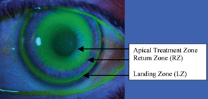
The optimal fit and zone alignment of an ortho-k lens.
Ortho-k lenses iatrogenically induce corneal topographic sphericalization via constant pressure on the flat meridian and variable pressure on the steep meridian. A plateau is reached when the cornea becomes spherical secondary to applied uniform pressure, resulting in central flattening and peripheral steepening.26-28
Today, several companies manufacture ortho-k lenses. In June 2002, Paragon Vision Sciences’ Corneal Refractive Therapy lens was the first ortho-k design to gain FDA approval for overnight wear. Subsequently, Euclid Systems’ Emerald lens secured FDA approval in 2004 (Bausch + Lomb purchased the lens design in 2005).
Ortho-k for Myopia Control
More than a decade ago, several eye care clinicians initially suggested that ortho-k lenses potentially could be used to control or even halt myopia progression. Then, in 2004, the results from the first report on ortho-k for myopia control––the Children’s Overnight Orthokeratology Investigation (COOKI) pilot study––were published.29 COOKI researchers evaluated refractive error, visual changes and ocular health for six months in myopic children who were fit with overnight ortho-k lenses. The researchers determined that overnight ortho-k was both a safe and effective treatment for curtailing myopia progression.
In 2005, data from the Longitudinal Orthokeratology Research in Children (LORIC) study indicated that ortho-k was effective at controlling childhood myopia.30 However, the researchers also determined that substantial anatomic variations among children can reduce the clinician’s ability to accurately predict final visual outcome before starting ortho-k therapy.30
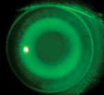
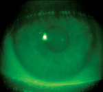
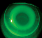
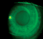
A) Optimally centered fit of an ortho-k lens. B) Cornea following optimally centered lens removal. Note virtually no trace of an impression ring. C) Superior-nasal decentered fit of an ortho-k lens. D) Impression ring following decentered lens removal.
Results from more recent clinical trials, such as the Stabilizing Myopia by Accelerated Reshaping Technique (SMART) study and the Corneal Reshaping and Yearly Observation of Myopia (CRAYON) study, have yielded additional information regarding the safety and efficacy of ortho-k for myopia control.31,32
SMART, a five-year study initiated in 2009, currently is evaluating the effect of ortho-k on myopia progression in 138 patients. At one-year follow-up, subjects wearing ortho-k lenses exhibited a mean progression of 0.00D, compared to an average of 0.50D in the control group.31
In the two-year CRAYON study, researchers confirmed that patients who were fitted with ortho-k lenses experienced significantly less annual change in axial length and vitreous chamber depth than patients fitted with soft contact lenses.32 These results confirmed data from previous studies by showing that ortho-k lenses slow the progression of corneal changes in myopic patients.32
Recently, there has been suggestion of low-level myopic orthokeratology with the use of a high-modulus silicone hydrogel lens intentionally worn in an everted position. The everted wear of a high-minus silicone hydrogel contact lens can induce alterations in corneal topography and subjective refraction. These refractive change range from plano to +1.75D sphere and +0.25D to +0.75D cylinder, but are unpredictable and vary from subject to subject.33,34
In one study, the mean apical topographic power change was 1.11D with slight corneal steepening in both meridians as well as 0.23mm of corneal flattening in the horizontal meridian and 0.27mm of corneal flattening in the vertical meridian.33 Additionally, corneal eccentricity decreased by an average of 0.65e.34 These results suggest that this ortho-k technique may be suitable for patients with very low myopia.
Safety Concerns and Side Effects
As the aforementioned studies suggest, ortho-k is a safe procedure as long as patients are monitored properly. As with all contact lens use, the two most common side effects that occur in patients with ortho-k lenses are corneal edema and staining. Other potential side effects include pain, redness, tearing, irritation, discharge, ocular abrasion or visual distortion.35-37 These usually are temporary conditions, especially if the lenses are removed promptly. However, clinicians must be particularly cautious to monitor for microbial lens binding of Pseudomonas aeruginosa.35-37 Instruct patients to report any pain, discomfort or visual compromise immediately.
As with any contact lens or refractive procedure, induced visual aberrations and distortions can occur and should be monitored using aberrometry. Following overnight ortho-k, one study uncovered significantly lower rates of mesopic contrast as well as an increased incidence of higher-order aberrations. In another study, researchers found that higher-order aberrations––especially spherical aberration and coma––had a short-term, but significant, increase following ortho-k.38 Fortunately, these visual effects usually are reversible within 72 hours of lens discontinuation.38
Other Discussion Points
• Residual cylinder. Ortho-k lenses are designed for individuals with low to moderate myopia (up to -6.00D) with or without astigmatism (up to -1.75D). But beware of higher cylindrical correction and possible residual cylinder—you may reduce the myopia but leave appreciable higher-order aberrations with uncorrected cylinder.
• Astigmatism. Even though astigmatism is addressed at low levels with traditional orthokeratology, Paragon Science is launching the dual-axis system to addresses higher cylinder corrections. In the traditional design, a lens fit on the flat meridian would de-center due to the differential between the flat and steep meridians. (Sometimes, this may be seen as an ortho-k island that is topographically similar to a LASIK island.) The dual axes have a spherical base curve with a unique peripheral system. This affords the two meridians varied depths within the return zone.39
• Full distance vs. monovision. In an adult patient, you can either set a goal of full-distance correction or monovision. In a full-distance correction, the patient will require reading glasses. If, however, the patient is fit with monovision and does not like the effect, simply adjust the base curve to push for a fuller distance or an intermediate distance correction.
In children, the full-distance correction will be required. However, do not abandon the concept of readers or even vision therapy.
Is Ortho-k Reversible?
As mentioned previously, the effects of ortho-k usually are temporary and reversible. This fundamental consideration often appeals to many patients who are intimidated by the lifelong effect of LASIK or PRK. Additionally, adult patients are more comfortable knowing that ortho-k lenses can be manipulated as needed to address presbyopia throughout the child’s lifetime.
In order to maintain the long-term refractive effect of ortho-k, however, the patient must wear a retainer lens. Again, similar to the use of a dental retainer in orthodontia to maintain teeth alignment, a retainer lens is worn to preserve the flattening effect and prevent further myopia progression.
Individual Patient Needs and Considerations for Ortho-k
* Toric and dual-axis ortho-k lenses are becoming more widely available.44
Overnight ortho-k lens wear compresses the corneal epithelium, forcing a reduction in central corneal thickness, without damaging the epithelial cells or the contiguous stromal tissue. Once the lens is removed, the cornea does not simply bounce back like a “top hat,” but rather slowly returns to its natural, pre-treatment state. The compressed effect should persist for at least 12 to 15 hours, while the patient is awake (in some cases, the effect may last two to three days). Then, to maintain the compressed shape, the retainer lens must be placed on the eye once again during the nighttime hours.
In my clinic, we instruct the patient to wear the retainer lens each night for at least 30 days. After 30 days, we ask the patient not to wear the retainer lens for one night to determine the longevity of its effect. We explain that they may lose some of the effect throughout the second day; however, the accommodative loss will be based on subjective impression. If the patient appreciates a more rapid blur, we suggest that the retainer should be worn each night (or even every other night), as long as he or she retains comfortable and functional vision. Anecdotally, I have found that some patients have been very successful without the retainer for three full days, while others simply prefer to use the lens each evening. No matter the case, it is best to reassure your patients that, if they miss a night of retainer wear, they will not lose the effect.
The reversibility of ortho-k’s effect varies per individual; however, patients generally notice regressive changes within 24 to 72 hours. Furthermore, regression is not linear and thus not absolutely predictable. Refractive error, patient age and subjective tolerance are the determining factors of appreciable visual regression.40 And, because of the subjective considerations, perceived regression is not always consistent with objective refractive and topographic findings. Keep in mind, however, that patients with higher levels of baseline myopia seem to be more likely to experience end-of-day regression.40,41
Prepare the Patient
Keep in mind that some prospective ortho-k patients never wore contact lenses before. In my office, we ask our novice patients to participate in a trial of daily disposable or silicone hydrogel lenses. This will teach the patient how to insert and remove the lenses properly as well as how to handle the lenses and care products.
Furthermore, this contact lens trial also gives us an avenue for myopia correction if, by chance, the patient is not successful with ortho-k lenses. (As a side note, our office recommends a contact lens trial during refractive surgical preparation. We prescribe lenses with the targeted refractive correction, or lesser, to facilitate visual education as well as provide a realistic illustration of the anticipated surgical outcome.)
The patient should be educated that, as with any new fit, there is lens awareness at first (the sensation often will become less noticeable after sleeping in the lenses). Also, the initial lens fitting can be enhanced with a diluted dose of proparacaine, if needed. Fortunately, ortho-k lenses are very comfortable due to their large size and contour.
Guiding Patient Expectations
The modern technology of the corneal reshaping lenses allows for immediate appreciation of the myopic reduction; however, it takes extensive training time for the cornea to hold its new shape consistently. During the first weeks, the lenses should be worn every night. Thus, it is important to establish a proper and realistic timeline for the patient. Equally important: Do not over-promise visual results.
When discussing expectations with younger patients, it is important to respect parental input––but not at the expense of the child. If the child is uncomfortable with the concept of contact lens wear or corneal reshaping, do not let the parent force ortho-k on the child. For ortho-k to be successful, the child (and/or parent) must genuinely believe in the corrective process and conscientiously decide to remain compliant with lens wear.
As suggested by results from the LORIC study, each patient has unique visual needs as well individualized expectations and endpoint visual goals (see “ Individual Patient Needs and Considerations for Ortho-k."). In the first week, ortho-k lenses could be worn throughout the day (if tolerated by the patient) to accelerate the effect. However, this step may not be necessary or desirable for all patients.
Additionally, create an informed consent contract that formally outlines the fitting method and appropriate aftercare. This contract should define all concerns and precautions of contact lens wear, lens care product prescriptions, patient responsibilities, information about the exchange of lenses and warranties, replacement policies and your contact information for emergency care. The contract also should itemize the cost of professional fees and associated materials as well as detail all financial arrangements.
On the first day of lens wear, the patient will need to be seen in the morning and afternoon to determine if the lens settled into the correct position after sleep and to monitor for regression. You should also schedule several follow-up appointments to monitor for refractive and topographic changes (see “ An Ideal Short-term Follow-up Schedule.").
After approximately one month, the patient can attempt to discontinue lens wear for 48 to 72 hours to determine the longevity of the effect and build confidence in the procedure. This step also will help the patient to decide if he or she wants to wear the retainer lens every night or every other night.
Generally, our office will use this follow-up schedule with all ortho-k patients.
An Ideal Short-term Follow-up Schedule
Financial Costs of Ortho-k
The cost of ortho-k is unique to each practice, because of different lens models and varied professional fees. Also, regional pricing differences occur based on localized economics and patient demand. Some offices will arrange financing through an external company or have an arrangement policy that should be detailed in the informed consent contract.
Without question, however, ortho-k is a unique practice differentiator. Not all offices offer this service, leaving a window of opportunity for an individual practice to expand on the service or even consider comanagement with area colleagues.
Keep in mind that not all patients will be successful with ortho-k, so you may wish to offer an “easy out” for unsuccessful cases that poses minimal financial risk for the patient.
Offering ortho-k in your practice is an excellent opportunity to better serve the next wave of young myopes. As with LASIK, the popularity of ortho-k has dramatically decreased over the past several years––even in geographic areas once thought to be “recession proof.”42,43 While there is little to no published literature on the key financial data associated with orthokeratology, overall demand for the procedure would most likely follow refractive surgery trends. Nonetheless, it is logical to believe that as the eye care market and global economy strengthen, so too will the interest in ortho-k.
Dr. Daniels is in private practice in Hopewell and Lambertville, N.J. He also is an assistant clinical professor at Salus University in Elkins Park, Pa., and an external education preceptor for New England College of Optometry in Boston. He has served on the advisory panels for Tracey Technologies, WaveTouch Technologies, Sauflon Pharmaceuticals, VMax Technology, Hydrogel Vision, SynergEyes, CooperVision, ScienceBased Health and QSpex, and has participated in sponsored research for Allergan, Alcon, CooperVision, Vistakon and SynergEyes.
1. Mutti DO, Jones LA, Moeschberger ML, Zadnik K. AC/A ratio, age, and refractive error in children. Invest Ophthalmol Vis Sci. 2000 Aug;41(9):2469-78.
2. Mutti DO, Sholtz RI, Friedman NE, Zadnik K. Peripheral refraction and ocular shape in children. Invest Ophthalmol Vis Sci. 2000 Apr;41(5):1022-30.
3. Zadnik K, Mutti DO, Friedman NE, et al. Ocular predictors of the onset of juvenile myopia. Invest Ophthalmol Vis Sci. 1999 Aug;40(9):1936-43.
4. Zadnik K, Satariano WA, Mutti DO, et al. The effect of parental history of myopia on children’s eye size. JAMA. 1994 May 4;271(17):1323-7.
5. Zadnik K, Mutti DO, Friedman NE, Adams AJ. Initial cross-sectional results from the Orinda Longitudinal Study of Myopia. Optom Vis Sci. 1993 Sep;70(9):750-8.
6. Zadnik K, Manny RE, Yu JA, et al. Ocular component data in schoolchildren as a function of age and gender. Optom Vis Sci. 2003 Mar;80(3):226-36.
7. Rose KA, Morgan IG, Ip J, et al. Outdoor activity reduces the prevalence of myopia in children. Ophthalmology. 2008 Aug;115(8):1279-85. Epub 2008 Feb 21.
8. Saw SM. A synopsis of the prevalence rates and environmental risk factors for myopia. Clin Exp Optom. 2003 Sep;86(5):289-94.
9. Goldschmidt E. The mystery of myopia. Acta Ophthalmol Scand. 2003 Oct;81(5):431-6.
10. Vitale S, Sperduto RD, Ferris FL. Increased prevalence of myopia in the United States between 1971–1972 and 1999–2004. Arch Ophthalmol. 2009 Dec;127(12):1632-9.
11. Lin LL, Shih YF, Tsai CB, et al. Epidemiologic study of ocular refraction among schoolchildren in Taiwan in 1995. Optom Vis Sci. 1999 May;76(5):275-81.
12. Mitchell P, Hourihan F, Sandbach J, Wang JJ. The relationship between glaucoma and myopia: the Blue Mountains Eye Study. Ophthalmology. 1999 Oct;106(10):2010-5.
13. Kerns RL. Contact lens control of myopia. Am J Optom Physiol Opt. 1981 Jul;58(7):541-5.
14. Kemmetmüller H. Can myopia be influenced by the wearing of contact lenses? Klin Monbl Augenheilkd. 1976 Jan;168(1):10-23.
15. Shapiro EI, Kivaev AA, Kazakevich BG. Use of contact lenses in progressive myopia. Vestn Oftalmol. 1990 Sep-Oct;106(5):30-3.
16. The Orthokeratology Academy of America. What is Orthokeratology? Available at: http://okglobal.org/home.html (accessed June 26, 2012).
17. Chua WH, Balakrishnan V, Chan YH, et al. Atropine for the treatment of childhood myopia. Ophthalmology. Dec;113(12):2285-91.
18. Walline JJ, Lindsley K, Vedula SS, et al. Interventions to slow progression of myopia in children. Cochrane Database Syst Rev. 2011 Dec 7;12:CD004916.
19. Ganesan P, Wildsoet CF. Pharmaceutical intervention for myopia control. Expert Rev Ophthalmol. 2010 Dec 1;5(6):759-87.
20. Siatkowski RM, Cotter SA, Crockett RS, et al. Two-year multicenter, randomized, double-masked, placebo-controlled, parallel safety and efficacy study of 2% pirenzepine ophthalmic gel in children with myopia. J AAPOS. 2008 Aug;12(4):332-9.
21. Trier K, Munk Ribel-Madsen S, Cui D, Brøgger Christensen S. Systemic 7-methylxanthine in retarding axial eye growth and myopia progression: a 36-month pilot study. J Ocul Biol Dis Infor. 2008 Dec;1(2-4):85-93.
22. Wei ML, Liu JP, Li N, Liu M. Acupuncture for slowing the progression of myopia in children and adolescents. Cochrane Database Syst Rev. 2011 Sep 7;9:CD007842.
23. Ciuffreda KJ, Ordonez X. Vision therapy to reduce abnormal nearwork-induced transient myopia. Optom Vis Sci. 1998 May;75(5):311-5.
24. Vasudevan B, Ciuffreda KJ, Ludlam DP. Accommodative training to reduce nearwork-induced transient myopia. Optom Vis Sci. 2009 Nov;86(11):1287-94.
25. Gwiazda J, Hyman L, Hussein M, et al. A randomized clinical trial of progressive addition lenses versus single vision lenses on the progression of myopia in children. Invest Ophthalmol Vis Sci. 2003 Apr;44(4):1492-500.
26. Ziff SL. Orthokeratology 1. J Am Optom Assoc. 1968 Feb;39(2):143-7 contd.
27. Nolan JA. Approach to orthokeratology. J Am Optom Assoc. 1969 Mar;40(3):303-5.
28. Binder PS, May CH, Grant SC. An evaluation of orthokeratology. Ophthalmology. 1980 Aug;87(8):729-44.
29. Walline JJ, Rah MJ, Jones LA. The Children’s Overnight Orthokeratology Investigation (COOKI) pilot study. Optom Vis Sci. 2004 Jun;81(6):407-13.
30. Cho P, Cheung SW, Edwards M. The longitudinal orthokeratology research in children (LORIC) in Hong Kong: a pilot study on refractive changes and myopic control. Curr Eye Res. 2005 Jan;30(1):71-80.
31. Global OK-Vision. Orthokeratology Procedure. Available at:
www.govlenses.com/orthokeratology/procedure.html (accessed June 26, 2012).
32. Walline JJ, Jones LA, Sinnott LT. Corneal reshaping and myopia progression. Br J Ophthalmol. 2009 Sep;93(9):1181-5.
33. Bogert A. Silicone Hydrogel Orthokeratology for the Correction of Low Myopia Thesis presented at University of Applied Sciences Aalen, Germany. Available at:
http://opus.bsz-bw.de/hsaa/volltexte/2010/2/pdf/Silicone_Hydrogel_Orthkeratology_for_the_Correction_of_Low_M.pdf (accessed June 26, 2012).
34. Mountford J. Unintended orthokeratology effect of silicone hydrogels on hypermetropic patients. Available at:
www.siliconehydrogels.org/featured_review/featured_review_aug_03.asp (accessed June 26, 2012).
35. Ladage PM, Yamamoto N, Robertson DM, et al. Pseudomonas aeruginosa corneal binding after 24-hour orthokeratology lens wear. Eye Contact Lens. 2004 Jul;30(3):173-8.
36. Young AL, Leung AT, Cheung EY, et al. Orthokeratology lens-related Pseudomonas aeruginosa infectious keratitis. Cornea. 2003 Apr;22(3):265-6.
37. Choo JD, Holden BA, Papas EB, Willcox MD. Adhesion of Pseudomonas aeruginosa to orthokeratology and alignment lenses. Optom Vis Sci. 2009 Feb;86(2):93-7.
38. Stillitano IG, Chalita MR, Schor P, et al. Corneal changes and wavefront analysis after orthokeratology fitting test. Am J Ophthalmol. 2007 Sep;144(3):378-86.
39. Herzberg C, Legerton J. The advancing specialty of CRT. RCCL. 2011 Apr;147(3):20-3.
40. Gardiner HK, Leong MA, Gundel RE. Quantifying regression with orthokeratology. Contact Lens Spec. Available at:
www.clspectrum.com/articleviewer.aspx?articleID=12892 (accessed June 26, 2012).
41. Chan B, Cho P, Cheung SW. Orthokeratology practice in children in a university clinic in Hong. Clin Exp Optom. 2008 Sep;91(5):453-60.
42. Market Scope. Quarterly Updates on the U.S. Refractive Market.Q4-2009 U.S. Refractive Update. Available at:
www.market-scope.com/market_reports/refractive_reports.html (accessed June 26, 2012).
43. Laser Eye Surgery Industry Growth and the Economy. LASIK Industry Growth and the Economy. Available at:
www.laser-eye-surgery-statistics.com/lasik-economy-lasik-industry-growth.php (accessed June 26, 2012).
44. Baertschi M. Correction of high astigmatism with orthokeratology. Paper presented at the Global Orthokeratology Symposium. July 28-31, 2005; Chicago.

