 |
I have had the pleasure of taking a walk back in time by looking over a few glaucoma-focused articles published as far back as the 1930s. It’s been an interesting journey. Some things, in particular technological advances, have skyrocketed optometry and its role in managing glaucoma. Other topics have changed very little.
Allow me to be your tour guide as we step back in time to glaucoma treatments of yesteryear.
A Foot in the Door
In 1937, a lecturer at the University of California, Berkeley, penned two columns that appeared in Review relating to perimetry and the study of the visual fields.1,2 His first words echo that most of the basics of perimetry and the visual field are based on neuronal and retinal anatomical considerations established in the preceding 40 years.1 That tenet still exists. Visual field loss—whether it occurs from glaucomatous, neuro-ophthalmic or retinal origins—is always correlated with anatomic foci of aberrant or damaged tissue. While perimetric techniques have changed since that time, the interpretation and correlation to the structural etiology of the field defect remains the same: we examine the field defect and determine its anatomical source by essentially dividing visual field loss into three categories: prechiasmal field loss, chiasmal field loss and retrochiasmal field loss.
While the topic of these columns pertained to the optometric use of visual field testing devices, it centered on glaucomatous field loss. Interestingly, a portion of the column was spent describing the construction of a tangent screen, its simple design and the simple materials used. The slant was to stimulate an interest among optometrists that, with some easily obtained materials, one could construct a tangent screen and begin measuring visual fields. Many ODs today only think of automated perimetry. But, back in the day, some of us were trained on kinetic perimetry. Tangent screens were elegant, simple and, with proper technique, able to help identify glaucomatous field defects. However, they were labor intensive. Subsequently, Goldmann perimetry was developed, and used for many years clinically. While I was at the Pennsylvania College of Optometry, we had the first exposure to the newest form of perimetry: the fully automated Humphrey perimeter. It was about the size of a refrigerator, but it heralded the era of automated perimetry. One fundamental difference between tangent screens (and Goldmann perimeters) versus these new Humphrey perimeters, was stimulus presentation. While I did not use a tangent screen (I’m old, but not that old!), I did regularly use a Goldmann perimeter. These perimeters presented a kinetic stimulus to the patient. The mechanics from the patient’s point of view were the same: they pushed a button when they “saw the light.” Goldmann perimeters could be deadly accurate, but one needed a superb technician driving the test to obtain that accurate field study. Needless to say, standardization of testing parameters became the norm, along with less dependence on technician abilities. Thus, the huge switch to what we know today: standard automated perimetry (SAP). Of course, typical SAP strategies use a stationary fixed stimulus rather than a kinetic stimulus, but the end result, ultimately, is the same identification of a visual field defect.
 |
| A look inside a 1937 Optical Journal and Review of Optometry article on glaucoma. Click to enlarge. |
One can go back today and read Hobson’s columns, and gain a good and rather accurate understanding of the anatomical correlates to visual field defects. One of the most useful graphics in understanding visual fields, as seen in his columns, is the illustration of the field of vision as a ‘hill of vision’, or sometimes called an island of vision in a sea of blindness. That graphic is a great tool in understanding the relationship between sensitivity of retinal fields, and the relationship between sensitivities and distances from the central retina. It worked then, and it still works today. And it is the basis of visual field testing just as much today as visual field testing back in the early 1900s.
An Invitation
In the early 1940s, Harold Noyes, MD, penned a column that appeared in The Optical Journal, for optometrists, outlining the various types of glaucoma known at that time.3 The intent of his column was to educate optometrists about glaucoma, in the hopes that a well-trained and well-equipped optometrist could detect the disease and subsequently refer the patient for appropriate care. Remember, this was well before any optometrists could legally use diagnostic or anesthetics drops. I’m sure it was a bold move at the time and I would guess he caught some heat for discussing glaucoma in an optometric journal. But he must have realized an inevitable truth: the majority of patients at the time sought refractive care and, therefore, ended up seeing an OD for their visual needs. With the OD as the entry point into the eye care system, he realized that educating this group of professionals was important in identifying those individuals who had glaucoma. Sure, ODs of this time could not treat the disease, but at least they could facilitate appropriate care for the patient.His review of the types of glaucoma was fairly accurate, though the methodology of examining the patients was somewhat limited by the technology at the time. He pointed out that the majority of cases of glaucoma fell into the category of what we today call primary “open angle glaucoma,” with insidious and asymptomatic onset.3 He also discussed angle closure glaucoma, which he called “chronic congestive glaucoma,” as well as other cases of secondary glaucomas related to retinal vascular diseases, inflammatory diseases and orbital diseases.3 One comment in the body of the piece speaks volumes to the technology at the time. He references that many cases of glaucoma initially labeled “simple glaucoma” may become recharacterized as a separate, different type of glaucoma with the use of a slit lamp to examine the eye and finding subtle differences and signs.3 Imagine that? Using a slit lamp to aid in the diagnosis of glaucoma!
Joining the Diagnostic Team
By the early ’50s, doctors recognized glaucoma was more prevalent than initially believed and that, because optometrists were often the first providers patients saw, it was incumbent upon us to become versed in identifying glaucoma patients. It was time for ODs to become a part of the diagnostic team. Interestingly, one of the columns from 1950 references the hearings on Capitol Hill for ODs to become a part of the outpatient care clinics in the Veterans Administration (VA) system, and how the diagnosis of glaucoma was used in arguments against ODs providing care. Sound familiar?
In 1951, a column titled “The Optometric Aspects of Glaucoma,” described examination techniques needed to identify the disease.4 Schiotz tonometry was a must, according to the article.4 Corneas should be clear and evaluation of the anterior segment should at least be accomplished with a plus condensing lens and a goose neck lamp.4 Finger palpation of the globes was recommended, too.4 Ophthalmoscopic findings consistent with glaucoma were discussed, as were gonioscopic findings.4 Remember, optometrists were still restricted from using diagnostic pharmaceutical agents in every state. However, the education process had begun.
In the world of optometric legislation, there is a wise saying: educate before legislate. Needless to say, this means that prior to legislating expanded OD licensure, it is incumbent upon the OD to be educated in a particular area or set of procedures. So, the wheels began to turn, gradually, back then. In 1952, Richard Struhl, OD, asked a basic question: was it not time for ODs to begin using “simple drugs for the detection and diagnosis of various eye diseases?”5 He quickly followed that up with a comment that these drugs should be nontherapeutic, but that’s how change happens. Slowly, over years.
Getting up to Speed
In the 1970s, optometric diagnostic and therapeutic laws started to pick up steam across the country. In 1975, Marvin Smith, PhD, adapted a lecture in the pages of The Optical Journal and Review of Optometry that sought to debunk myths circulating about diagnostic drugs.6 It may seem quaint today to think simple mydriatics and cycloplegics could be at the heart of such controversy, but in 1975 the debated warranted a four-page rebuttal.
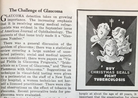 |
| A 1950 article, “The Challenge of Glaucoma,” runs beside an image ugring readers to “Buy Christmas Seals. Fight Tuberculousis.” |
The arguments that Dr. Smith was rebutting were the beginning of a decades long theme when optometric expansion was being criticized by those objecting to it, primarily the medical community. Comments like “topical anesthetics can cause shock, cardiac arrest and death,” “eye drops have caused death in infants,” and the classic “the public would be exposed to new risk from the use of these drugs [diagnostic agents] by optometrists,” and summarized by the other classic: “medical licensure is essential for the use of drugs.” To quote Dr. Smith: “The question arises as to why there were, and are, hard fought legislative and court battles between two groups of practitioners who both have the best interests of the public in mind? The answer to this question is indeed that there are myths and misinformation which pervade the use of diagnostic pharmaceutical agents in the eye.”6 Wow! The same tactics are used today by those opposed to optometric scope expansion.
The passage of DPA legislation allowed ODs to begin testing with these formulations and, in many ways, changed what it meant to be an optometrist. This was reflected in the pages of the rebranded Review of Optometry. For example, in 1986 virtually none of the magazine’s feature headlines even contained the word “glaucoma.” A year later, that changed drastically as Review launched a bimonthly “Glaucoma Series.”7 This was ground breaking coverage in an optometric journal. Glaucoma was a pervasive disease in the population, but was only treated by those with medical licenses, except in West Virginia and North Carolina where ODs were authorized to treat the condition. In the inaugural installment, Thomas Lewis, OD, covered the topic exquisitely. Interestingly, long before OHTS was even an idea, he noted that only about one in 10 ocular hypertensives actually went on to develop frank glaucoma. But the presence of that article in a national optometric publication was a part of a growing shift toward publishing more information in mainstream publications dealing with the optometric management of conditions involving other than refractive care.
In 1994, it launched its official “annual glaucoma report” issue, a tradition it keeps up today. And I have the honor and privilege of authoring the bimonthly “Glaucoma Grand Rounds” column.
Today
Imagine practicing without the prescriptive authority we now have. There is still work to be done. Just recently, New York State was prevented once again from allowing its ODs to prescribe oral pharmaceutical agents. When I graduated, only two states allowed ODs to prescribe TPAs, and Pennsylvania was not one of them. Growing up outside Philadelphia did not change the fact that I would not be able to use the majority of what I was taught. That’s how I ended up in North Carolina.
The wheels continue to turn. Often, more slowly than we want. But they need to continue to turn. Perhaps in the anniversary issue 100 years from now, some of the things that we currently do and consider cutting edge will be highlighted as quaint things ‘the old-timers’ used to do. I can only hope so.
|
1. Hobson JT. Method of procedure with Bjerrum screen. Part 1. Optical Journal-Review. December 1, 1937;64(23):16-8. 2. Hobson JT. Method of procedure with Bjerrum screen. Part 2. Optical Journal-Review. December 15, 1937;64(24):16-8. 3. Noyes HG. Glaucoma, its types and diagnostic symptoms. Optical Journal-Review. December 1, 1941;68(23):18-20. 4. Lopez RB. The optometric aspects of glaucoma. Optical Journal-Review. January 15, 1951;78(2):70-5. 5. Struhl R. Tonometry by optometrists. Optical Journal-Review. January 1, 1952;79(1):42. 6. Smith M. Myths and misinformation concerning use of diagnostic drugs. Optical Journal-Review. November 15, 1975;112(22):28-31. 7. Lewis T. Glaucoma: is your patient at risk? Review of Optometry. January, 1987;124(1):59-62. |
