 |
Q:
My patient has hemochromatosis and is interested in LASIK. There are no indications of ocular surface dryness, corneal disease or cataracts. Are there any known contraindications or concerns? Should we proceed with the surgery?
A:
Pigmented iron lines and deposits can occur in the corneas of healthy patients and patients with corneal pathologies, according to Vance Thompson, MD, director of refractive surgery at Vance Thompson Vision, and Mitch Ibach, OD, who practices at Vance Thompson Vision. They note that metallic foreign bodies can also leave rust rings and deposit iron in the cornea.
Hemochromatosis is a condition in which there is a buildup of iron in the body.1 In genetic hemochromatosis, the intestines absorb too much dietary iron, which then builds up throughout the body’s organs.2 Acquired hemochromatosis is usually secondary to anemia—excess breakdown of red blood cells—or over-absorption caused by iron transfusions.2 The human body can’t excrete excess iron, and systemic damage typically takes the form of toxic oxygen free radicals that then lead to oxidative damage.3
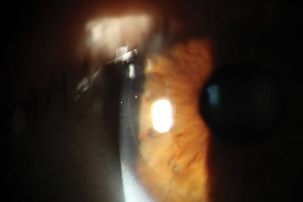 |
A clear, well-adhered LASIK flap seen one day postoperatively. Click image to enlarge. |
Setting Up For Success
Prior to corneal laser refractive surgery, eye doctors must establish that the patient’s tear film is healthy enough to aid in immediate healing and long-term refractive preservation, state Drs. Thompson and Ibach. Iron transport proteins lactoferrin and ferritin are both readily found in the tear film and may be dysregulated in hemochromatosis.3 Despite their iron-binding nature, over 90% of lactoferrin proteins remain unbound to iron in normal homeostasis.3 In rabbit corneas, unsaturated lactoferrin can protect against oxidative damage.3
Lactoferrin in its non-iron-bound form exhibits bactericidal properties and anti-biofilm activity.3 Analogous to lactoferrin, ferritin is also capable of warding off oxidative damage.3 In hemochromatosis patients, Drs. Thompson and Ibach say, it is possible that the positive attributes of these iron proteins are depleted but unclear if hemochromatosis has a negative effect on tear film quality or quantity.
The eye’s second main refractive structure is the crystalline lens, which has three refractive stages: optically clear with sufficient accommodation, optically clear with insufficient accommodation (presbyopia) and opaque/cloudy with insufficient accommodation (cataract). The pathogenic cause of cataracts is not fully understood, but evidence suggests oxidative damage plays a role.3 Drs. Thompson and Ibach highlight the importance of preoperatively educating hemochromatosis patients on the possibility of early cataract formation, which could shorten the refractive lifespan of corneal refractive surgery.
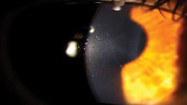 |
A PRK patient two weeks post-op. Click image to enlarge. |
Satisfaction rates for LASIK have risen to 98%, making this elective procedure one of the most successful.4 Drs. Thompson and Ibach emphasize that corneal refractive surgery, like any procedure, presents risks that patients must thoroughly understand ahead of time. They believe hemochromatosis patients can safely undergo laser refractive surgery and should expect outcomes on par with the published literature. In the United States, they add, FDA-approved options including LASIK, PRK and SMILE can all confidently be recommended to this patient.
| 1. Hemochromatosis.org. www.hemochromatosis.org/#overview. Accessed November 15, 2019. 2. Lazzaro DR, Lin K, Stevens JA. Corneal findings in hemochromatosis. Arch Ophthalmol. 1998;116(11):1531-2. 3. Loh A, Hadziahmetovic M, Dunaief JL. Iron homeostasis and eye disease. Biochim Biophys Acta. 2009;1790(7):637-49. 4. Donnenfeld ED. The best for LASIK. Presented at AAO Subspecialty Days; November 10-11, 2017; New Orleans. |
