Corneal injuries are often intensely painful and present the potential for ocular morbidity with vision loss. While the causes of abrasions and foreign bodies—and their effects on the eyes—are seemingly unlimited, the overall treatment goal is similar: minimize pain, maintain integrity of the globe, prevent corneal scarring and infection, and preserve visual acuity.
Patients with corneal trauma report redness, photophobia, decreased acuity (when the injury involves the visual axis) and varying degrees of pain. Because patients have different thresholds for pain, its presence or absence is not always helpful from a diagnostic perspective. However, management is often dictated by this factor.
Clinicians should always take a detailed history and document a patient’s visual acuity, which may have to be obtained while triaging them. Though the use of anesthetic drops may be necessary due to patient discomfort, clinicians should quickly assess the scope of the injury and attempt to check visual acuity, including pinhole, prior to the instillation of any medications.
Corneal Healing
When managing a corneal injury, it is helpful to remember these key points of the healing process:
• The cornea repairs by cell migration, proliferation and differentiation, followed by extracellular matrix remodeling.1
• Corneal epithelial healing relies on limbal stem cells and remodeling of the basement membrane.
• The corneal regenerative response to an abrasion is related to the size and depth of the wound. Small epithelial defects typically heal in 24 to 48 hours, whereas large defects may take significantly longer, particularly if the stroma is involved. Corneal edema may remain after the epithelium heals and may continue to cause a decrease in visual acuity until its resolution.
• For deeper injuries, the corneal stroma heals via the transformation of keratocytes to fibroblasts and myofibroblasts, which may result in opacification and scarring.1
• In the initial phase of healing, epithelial cells flatten, spread and move. Cellular and subcellular reorganization and migration of the epithelial cells also occurs at the wound edge.2
• Cell proliferation, necessary to heal large abrasions, begins approximately 24 hours after injury. Stem cells from the limbus give rise to transient amplifying cells (TAC), which migrate to heal the corneal defect and replenish the wounded area.3 The majority of the defect is covered by a single layer of epithelium that “slides” over the wound, with a normal thickness restored by proliferation and upward movement of cells from the basal layer.2 The wound healing is not complete until the newly regenerated epithelium has anchored firmly to the underlying connective tissue, which does not occur until the defect is completely covered. Although transient attachments are regularly formed and released during the cell migration process, formation of normal adhesions takes approximately six weeks.2
Management of Corneal Epithelial Defects
Corneal abrasions make up the majority of corneal injuries, and many treatment methods exist to resolve them without visual complications. Topical management typically involves:
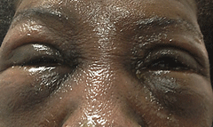
Case 1: The Exploding EggA 57-year-old black female presented urgently with eye swelling and facial burns from an “egg exploding in her face” after reheating it in the microwave, while in the shell. The patient reported initially to the emergency room and was diagnosed with facial and ocular burns and placed on erythromycin ointment for the eyes and Neosporin (neomycin/polymyxin B/bacitracin, Johnson & Johnson) ointment for her facial burns.
The following day, the patient reported to the clinic due to increasing lid edema and consistent ocular pain that was greater in the right eye than in the left. She was an established patient with a history of systemic lupus erythematosus and associated keratitis sicca. She was using Restasis (cyclosporine ophthalmic emulsion 0.05%, Allergan) OU BID and artificial tears for her dry eye condition. Systemically, she was taking Plaquenil (hydroxychloroquine, Sanofi-Aventis) 200mg BID, pantoprazole, omega-3 fatty acids, metoprolol, meloxicam, Imuran (azathioprine, Prometheus Laboratories), folic acid, aspirin, Rocaltrol (calcitriol, Roche) and Advair diskus by inhalation (fluticasone propionate and salmeterol inhalation powder, GlaxoSmithKline). The patient’s uncorrected visual acuity on initial examination was 20/100 OD and 20/80 OS. Clinical examination revealed blistering of the skin on the face as well as 4+ lid edema in both eyes (Figure 1). Her right cornea had a 6mm-by-5mm abrasion. In addition, there was 4+ SPK on both corneas. The anterior chamber was clear and well formed in both eyes. There was no evidence of foreign bodies or penetrating injuries. A bandage lens was placed on the patient’s right eye with some difficulty due to lid edema, and the patient noted immediate relief. She was instructed to discontinue the erythromycin ointment and was prescribed prophylactic moxifloxacin OD TID and artificial tears every hour in both eyes. The patient returned the following day and reported feeling significantly better. The bandage lens was moisturized with artificial tears and carefully removed. The corneal epithelial defect was significantly improved with a 1mm by 0.5mm defect remaining. A bandage lens was no longer needed, and the patient was advised to continue with her topical drops. The patient returned two days later, and her uncorrected visual acuity was 20/20 OD and 20/25+ OS. Her eyes were white and the edema had nearly resolved. The abrasion was healed, though there was still a 3+ SPK in both eyes secondary to her keratitis sicca. The patient was instructed to discontinue her moxifloxacin, continue artificial tears and resume her Restasis OU BID. The patient was seen one week later and her facial burns had nearly healed with some pigment abnormalities remaining and her keratitis sicca persists. |
• Antibiotics. This is administered as a prophylactic measure, and several options can achieve similar efficacies. Ultimately, antibiotic choice is often based upon physician preference. Fourth-generation fluoroquinolones have a wide spectrum of coverage, are less toxic than some traditionally used medications and are used regularly for prophylaxis in surgical patients.4-7 Other acceptable medications include Polytrim (trimethoprim/polymyxin B ophthalmic solution, Allergan), aminoglycosides (e.g., gentamycin or tobramycin) or less desirable early-generation fluoroquinolones such as ciprofloxacin and ofloxacin.
• Artificial tears. These are used to flush away antigenic material, promote epithelial repair and provide relief from discomfort.
• A cycloplegic agent. This will reduce secondary inflammation and uncomfortable ciliary spasm. It may be necessary to remove any retained foreign material or debride the loose epithelial edges to improve the healing process.
Small Abrasions
Lesions without significant loss of epithelial tissue generally heal well and quite quickly in the absence of
treatment. If a patient has minimal to no pain, an artificial tear with a prophylactic antibiotic is typically adequate. Advise patients to rest and reassure them that they will likely feel significantly better in the morning, given the minor nature of their injuries.
Large Corneal Abrasions
For patients with an abrasion affecting 25% to 50% of the cornea, prophylactic antibiotic, preservative-free artificial tears every hour and in-office cycloplegia (e.g., homatropine 5%) are generally adequate. Often it is difficult for the patient to obtain the cycloplegic medication at a pharmacy. However, since the majority of the wound will be repaired within 24 to 48 hours, in-office administration is often sufficient, especially if the patient is seen on a daily basis.
The largest corneal abrasions—affecting greater than 50% of the cornea—may take longer to heal and cause the patient significant pain. Over-the-counter analgesics or prescription medication, such as acetaminophen with codeine or hydrocodone, may be necessary for a short period of time.
These abrasions are often associated with stromal folds from edema. Once the epithelium is eroded, fluid will readily migrate into the cornea. The edema, once accumulated, will not clear until the epithelium completely regenerates, which may take as long or longer to resolve than the epithelial defect—the defect may take two weeks to re-epithelialize, while the edema may last for up to six weeks.8 Large abrasions are treated with prophylactic antibiotics, copious artificial tears every hour and a cycloplegic agent. A slightly stronger cycloplegic such as atropine 1% may be necessary. If the patient is in significant pain, an NSAID or prescription oral analgesic may be used. Topical hyperosmotics may be helpful for resolving corneal edema and promoting tighter attachments of the epithelial cells to the basement membrane.
Bandage contact lenses can be very helpful for patients with significant pain from large abrasions. However, tight bandage lenses or materials with low oxygen permeability may worsen corneal edema. For this reason, only high Dk lenses should be used for this purpose.9 The newly formed epithelial attachments are very weak and may be pulled off easily when removing the bandage lens. To avoid this, float the contact lens in solution or artificial tears first to be certain it is loose before removing. Bandage contact lenses should be replaced at each follow up as necessary for pain management, as the cornea will need to be assessed and the patient will be using antibiotic drops over the lens. It is important to differentiate an epithelial defect from a contact lens-related corneal infiltrate. Bandage contact lenses should not be used to treat epithelial defects associated with contact lens wear.
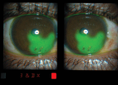
Case 2: Would You Like Bacon With that Egg?A 35-year-old black female presented emergently with severe pain in the right eye. She reported feeling intense pain that started suddenly while cooking breakfast. She believed she was splashed with hot oil. The patient’s visual acuity was 20/200 OD.
Clinical examination revealed a corneal abrasion as well as foreign body particles that appeared to be small pieces of bacon (Figure 2). The anterior chamber was clear and well formed. The eye was irrigated, the foreign bodies removed with forceps and the loose corneal epithelium debrided. The patient was treated with a bandage contact lens, a prophylactic antibiotic and artificial tears every 30 minutes to an hour. The patient returned the following day feeling significantly better, with the abrasion approximately 75% resolved. The bandage lens was replaced and her topical medications continued. When the patient returned 48 hours following the accident, her cornea was completely healed. Topical antibiotics were discontinued and she continued with artificial tears for comfort as needed. |
Topical ointments are commonly used to treat epithelial injuries. However, ointments are not sustained-release medications and thus provide little benefit. Further, while topical ointments may provide some lubrication, they may blur vision.
Any drug placed on the surface of the eye is in some way toxic and may interfere with re-epithelialization. Therefore, the “less is more” philosophy is a prudent choice when treating corneal abrasions.
A secondary infection from an abrasion is rare today due to the routine use of prophylactic topical antibiotics. Though studies on the conversion of corneal trauma to infectious keratitis or abscess are scant, ocular trauma is considered one of the most common causes of secondary infections following contact lens wear.10-12 One study revealed that as much as 15% of the bacterial keratitis in younger patients resulted from trauma, with even higher rates in rural areas, and is the number one cause in developing countries.12 When it does occur, an infectious ulcer or abscess on the cornea can have devastating consequences to the patient, potentially leading to perforation and vision loss. Therefore, topical antibiotics should always be used if possible.
Recurrent corneal erosion (RCE) may occur following a re-epithelialized abrasion—within days after the initial injury, or potentially months to years later in chronic cases due to poor anchoring of the epithelial cells to the basement membrane. The dry state of the eye’s surface when sleeping, combined with the weak attachments to the basement membrane, cause the epithelial tissue to lift off with the lids. RCEs are more common following injury in patients with basement membrane dystrophy.13 Typically, patients experience pain upon wakening similar to that of a small abrasion. RCEs are initially treated the same as a small abrasion, though repeat episodes may require additional procedures such as anterior stromal puncture, diamond burr debridement or laser resurfacing. There is also anecdotal evidence supporting the use of amniotic membrane tissue as a new treatment method for severe RCE.14-17
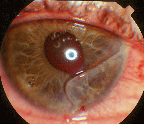
Case 3: Home Repair NightmareA 21-year-old plumber came in for an emergency visit after hitting himself with the blunt end of a screwdriver. He noted fluid running down his cheek and blurry vision. He also noted a “loose piece of skin” on his eye, which he attempted to remove. He was taking acetaminophen for discomfort.
Upon examination the patient was in extreme pain and had reduced visual acuity. A linear corneal defect was noted as well as an irregular pupil and bubbles in the anterior chamber. Additional testing revealed a positive Seidel’s test (Figure 3). The patient was diagnosed with a corneal laceration, and a shield was placed over the eye. He was referred for immediate surgical repair. |
A mild to moderate anterior chamber reaction may be associated with corneal injury. This inflammation generally resolves as the abrasion heals, but can respond well to a cycloplegic agent alone.
Thermal Burn Abrasions
Ocular thermal burns can occur upon exposure to flame, scalding liquid, blast injury or handheld objects such as curling irons and cigarettes. Injury associated with thermal burns is estimated to be between 7.5% and 27% of ocular trauma cases, with periocular damage more common than ocular surface damage due to the blink reflex and Bell’s phenomenon.18-20
In cases of severe burns, diagnosis and management may be delayed due to the critical nature of the condition. However, patients with superficial and partial thickness injuries may present to the optometric practice acutely for initial care.
Management of a thermal corneal defect is similar to that of an abrasion. Many traumatic epithelial defects require debridement to promote quicker healing, and often the necrotic white epithelium can simply be wiped off with a swab or Weck-Cel (Beaver Visitec). Prophylactic antibiotics are indicated at the minimum therapeutic dose to prevent bacterial colonization. Cycloplegics may be used if the patient is in pain or an inflammatory reaction has started, and may be instilled in-office since thermal corneal thermal defects are likely to heal quickly. Homatropine 2% or 5% as well as scopolamine 0.25% are appropriate. Preservative-free artificial tears help to aid in the re-epithelialization process and should be used frequently—as often as every 30 to 60 minutes. Topical steroids may be used on follow-up if secondary inflammation persists. Oral analgesics may also be used as needed for patients experiencing severe pain from large thermal abrasions. Bandage lenses are helpful; carefully remove and replace them on every follow-up.
Severe corneal and conjunctival thermal damage may cause symblepharon, corneal ulceration, perforation, scarring, neovascularization or limbal stem cell damage. These patients require amniotic membrane placement or surgical consultation for tarsorrhaphy, symblepharon ring and/or limbal stem cell transplantation.20-21 In some cases, oral doxycycline or ascorbic acid can be used for collagen synthesis.19 Scleral lens vaulting for corneal coverage is also used while awaiting surgical repair for severe thermal injuries.19
Chemical Burn Abrasions
Acids with pHs less than four and bases with pHs greater than 10 induce burns. Acidic compounds bind with tissue proteins and create their own barrier, whereas alkaline compounds saponify and “melt” fatty tissues, causing further penetration, leading to injuries more severe than acids.
The diagnostic signs of burns are redness or blanching of the conjunctiva, edema and burns to the skin or lids, anterior segment inflammation and corneal staining or haze. Corneal haze is correlated with the severity of the burn and the prognosis, according to the Roper-Hall (Ballen) classification: 22
• Grade I: no corneal haze and a good prognosis
• Grade II: some corneal haze but iris details are visible with a good prognosis
• Grade III: total epithelial loss with stromal haze obscuring iris details and a guarded prognosis
• Grade IV: an opaque cornea and no iris or pupil details visible and a poor prognosis
A white, blanched eye indicates destruction of the vessels and eventual tissue necrosis. Chemical insult may also damage the epithelial stem cells located at the limbus, resulting in delayed healing, conjunctivalization, corneal vascularization, conjunctival epithelial in-growth and opacification.8 It may also result in secondary glaucoma and requires careful monitoring of IOP.
Severe burns may require penetrating keratoplasty (PKP) with limbal stem cell transplant, amniotic membrane transplant or oral mucosal cell sheet transplant.23-26 Indications for surgical intervention involve chronic pain, severe ischemia, destruction of limbal stem cells and corneal destruction.
The initial management of any chemical burn is the same: copious irrigation. Patients calling to report chemical trauma should be advised to irrigate their eyes for 30 minutes prior to presenting to the office. When the patient arrives, use a litmus test to determine if the substance was acidic or alkaline. All patients should be irrigated upon arrival, including sweeping of the fornices for any particulate material. The severity of acid burns is typically clinically observable the day of the exam. In the case of alkaline burns, a patient’s presentation may worsen over a 24-hour period; they should be monitored very closely.
The goal of treating chemical burns is to control the inflammation of the underlying corneal stroma, preserve the limbal vasculature and restore the limbal stem cells. Managing chemical burns involves topical antibiotics, topical steroids with careful monitoring, artificial tears and oral analgesics as necessary. Take care when using bandage contact lenses with chemical burns, as any residual chemical remaining in the eye may impregnate the lens material and lengthen the contact with the eye.
Patients with mild acidic chemical burns generally respond well to preservative-free artificial tears every hour, prophylactic antibiotic drops and possibly in-office cycloplegia if the patient is in discomfort. Moderate to severe acid burns may require the addition of a mild to moderate strength topical steroid, such as fluorometholone or Lotemax (loteprednol, Bausch + Lomb) QID, or possibly prednisolone acetate 1.0%.
Topical steroids play an important role in managing alkaline burns in particular. These injuries have a biphasic pattern: the initial burn, then the secondary endothelial breakdown. Steroids are helpful in preventing the secondary breakdown and promoting endothelial repair.27 In addition, topical steroids prevent goblet cell loss and improve ocular surface health.28 Alkaline burns result in the release of collagenases and proteases, leading to corneoscleral melting.29 Topical steroids are useful for managing inflammation, but should be monitored very carefully. Collagenase inhibitors may be used in patients with severe corneal thinning who are at risk of perforation. Monitor these patients closely.
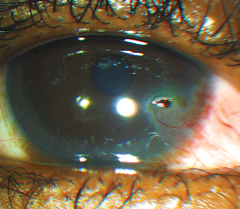 |
| Fig. 4. Foreign body of a portion of a bug. |
Ascorbate and citrate may be useful in severe alkaline burns. Researchers evaluated patients using topical prednisolone 0.5% along with topical ascorbate 10% and found no association with increase corneal melt if topical steroids were used until re-epithelialization.29
Foreign Body Removal
Any type of material can lodge in the eye, from bug wings to metal to superglue (Figure 4). Removal of any foreign body involves a thorough examination, assessment of the foreign body’s depth and location, and then the plan for its complete removal. Instruments that may be helpful include a spud, which may be used to remove the object and scrape adjacent tissue, and jeweler’s forceps. If the object is metallic, rust may form, necessitating the use of an Alger brush. Following removal of the foreign body, the patient will be left with an abrasion, which should then be managed accordingly.
Corneal Lacerations
Fortunately, full-thickness corneal lacerations are not as common as abrasions. However, when they occur, they can be associated with tremendous ocular morbidity. The classic sign of a corneal perforation is a positive Siedel test demonstrating leaking of the aqueous from the anterior chamber. Additional signs include bubbles in the anterior chamber, pupil irregularity, corectopia, iris prolapse, shallow or flat anterior chamber, hypotony and extrusion of ocular contents.
When treating a patient you suspect has suffered a perforation, remember that topical medications are not formulated for intraocular use and their effects cannot be predicted if they enter the globe, and placing them in an open globe may place the patient at risk for infection.31 If you suspect the cornea has been perforated, use only sterile products, such as saline and a fluorescein strip.
If an antibiotic is deemed necessary due to a delay in repair, though not sterile, moxifloxacin is not preserved and is often used directly in the anterior chamber during cataract surgery. Rather than coating the laceration with the strip, apply it in small amounts along the laceration to better control the response and distinguish how much of the laceration is a full-thickness penetration. If you are certain the cornea is perforated, place nothing in the eye. Anything placed in the eye in an attempt to help the pain or prevent infection may further contaminate the eye and encourage the patient to rub or wipe the eye, increasing the likelihood of uveal extrusion. Simply apply a rigid shield over the eye to prevent the patient from touching the eye and send them immediately to a specialist for repair. Educate the patient to avoid food and water, as they will likely require anesthesia.
Surgical repair may include tissue glue for partial-thickness lacerations or small perforations of 2mm or less.32-33 Bandage lenses may be used over the tissue glue for an additional barricade. Amniotic membranes are typically used more often in nontraumatic corneal defects, but may have a role in lacerations and used in conjunction with tissue glue.34 Sutures are most often used for full-thickness perforations. Often this may be accompanied with an air or gas (e.g., perfluoropropane) tamponade to prevent aqueous leakage.35 Following repair of the laceration and successful preservation of the integrity of the globe, grafting may be necessary for improvement in visual acuity. Corneal grafting is typically delayed for three months to improve primary repair success.36
In cases of open globe injury, be vigilant for increased inflammation and visual degradation, which may indicate endophthalmitis requiring referral to a retinal specialist.
Conclusion
Corneal injuries are common, painful clinical encounters in optometric practice that require quick, accurate decisions. When evaluating patients with corneal injuries, the first step is to determine the nature and mechanism of the injury—only then can we formulate proper management strategies. Patients with any corneal injury need to be closely monitored until the injury resolves or you have made an appropriate referral. Patients will be grateful for your assurance, care and confidence.
Dr. Vollmer is associate professor of optometry and director of residency programs for Nova Southeastern University. Her interests lie in primary care and ocular disease.
|
1. Ljubimov A, Saghizadeh M. Progress in corneal wound healing. Progress Ret and Eye Res. 2015;49:17-45. 2. Dua HS, Gomes JA, Singh A. Corneal epithelial wound healing. Br J Ophthal. 1994;78(5):401-8. 3. O’Sullivan F, Clynes M. Limbal stem cells, a review of their identification and culture for clinical use. Cytotech. 2007;53:101-6. 4. Alfonso E, Crider J. Ophthalmic infections and their anti-infective challenges. Surv of Ophthal. 2005;50(Suppl. 1):S1-6. 5. Burke JM, Bower KS, Vanroekel RC, et al. The effect of fourth generation fluoroquinolones gatifloxacin and moxifloxacin on epithelial healing following photorefractive keratectomy. Amer J Ophthal. 2005;140(1):83-7. 6. Schlech BA, Alfonso E. Overview of the potency of moxifloxacin ophthalmic solution 0.5% (Vigamox). Surv of Ophthal. 2005 Nov;50(Suppl. 1):S7-15. 7. Sclech BA, Blondeau J. Future of ophthalmic anti-infective therapy and the role of moxifloxacin ophthalmic solution 0.5% (Vigamox). Surv Ophthal. 2005 Nov;50(Suppl 1):S64-7. 8. Carlson EC, Wang IJ, Liu CY, et al. Altered KSPG expression by keratocytes following corneal injury. Molecular Vision 2003;9:615-23. 9. Foulks GN, Harvey T, Raj CV. Therapeutic contact lenses: the role of high Dk lenses. Ophthalmol Clin North Am. 2003 Sept;16(3):455-61. 10. Baklouti K, Ayachi M, Mhiri N, et al. Corneal abscess presumed to be of bacterial origin. Bull Soc Belge Ophtalmol. 2007;305:39-44. 11. Stefan C, Nenciu A. Post-traumatic bacterial keratitis- a microbiological prospective clinical study. Oftalmologia. 2006;50(3):118-22. 12. Bourcier T, Thomas F, Borderie V, et al. Bacterial keratitis: predisposing factors, clinical and microbiological review of 300 cases. Br J Ophthalmol. 2003;87(7):834-8. 13. Diez-Feijoo E, Duran, J. Optical coherence tomography findings in recurrent corneal erosion syndrome. Cornea. 2015 March;34(3):290-5. 14. Kordić R, Suić SP, Jandroković S, et al. Application of the amniotic membrane extract (AMX) for the persistent epithelial defect (PED) of the cornea. Coll Antropol. 2013;37(1):161-4. 15. Suri K, Kosker M, Raber I, et al. Sutureless Amniotic Membrane ProKera for Ocular Surface Disorders: Short-Term Results. Eye & Contact Lens: Science & Clinical Practice. 2013;39(5):341–7. 16. Yildiz E, Nurozler A, Ozkan A, et al. Amniotic membrane transplantation: indications and results. Eur J Ophthalmol. 2008;18(5):685-90. 17. Prabhasawat P, Tesavibul N, Komolsuradej W. Single and multilayer amniotic membrane transplantation for persistent corneal epithelial defect with and without stromal thinning and perforation. Br J Ophthalmol. 2001;85:1455-63. 18. Lin A, Petel N, Yoo D, et al. Management of ocular conditions in the burn unit: thermal and chemical burns and Stevens-Johnson Syndrome/Toxic Epidermal Necrolysis. J Burn Care & Res. 2011;32(5):547-60. 19. Czyz C, Kalwerisky K, Stacey A, et al. Initial treatment of ocular exposure and associated complication in severe periorbital thermal injuries. J Trauma Injury, Infection, and Critical Care. 2011;71:1455-59. 20. Bouchard C, Morno K, Perkins J, et al. Ocular complications of thermal injury: A 3-year retrospective. J Trauma Injury, Infection, and Critical Care. 2001;50:79-82. 21. Liang X, Zhiping L, Lin Y, et al. A modified symblepharon ring for sutureless amniotic membrane patch to treat acute ocular surface burns. J Burn Care & Research. 2012;33:32-8. 22. Roper-Hall MJ. Thermal and chemical burns. Trans Ophthalmol. Soc UK. 1965;85: 631-53. 23. Frucht-Pery J, Siganos CS, Solomon A, et al. Limbal cell autograft transplantation for severe ocular surface disorders. Graefes Arch Clin Exp Ophth. 1998 Aug;236(8):582-7. 24. Kruse FE. Stem cells and corneal epithelial regeneration. Eye. 1994;8(Pt 2):170-83. 25. Singh P, Tyagi M, Kumar Y, et al. Ocular chemical injuries and their management. Oman J Ophthalmol. 2013;6(2);83-6. 26. Fish R, Davidson R. Management of ocular thermal and chemical injuries, including amniotic membrane therapy. Curr Opin Ophthalmol. 2010(21);317-21. 27. Gomes JA, Romano A, Santos MS, Dua HS. Amniotic membrane use in ophthalmology. Curr Opin Ophthalmol. 2005 Aug;16(4):233-40. 28. Brent BD, Karcioglu ZA. Effect of topical corticosteroids on goblet cell density in an alkali burn model. Ann Ophthal. 1991 June; 23(6):221-3. 29. Davis AR, Ali QK, Aclimandos WA, Hunter PA. Topical steroid use in the treatment of ocular alkali burns. Br J Ophthalmol. 1997 Sep;81(9):732-4. 30. Kabat A, Sowka J. Cornea Atlas: Part III: From abrasions to burns. How to manage corneal injuries. Review of Optometry. Oct 1999. 31. Chung JH, Paek SM, Choi JJ, et al. Effect of topically applied 0.1% dexamethasone on endothelial healing and aqueous composition during the repair process of rabbit corneal alkali wound. Curr Eye Res. 1999;18(2):110-6. 32. Siatiri H, Moghimi S, Malihi M, Khodabande A. Use of sealant (HFG) in corneal perforations. Cornea. 2008;27(9):988-91. 33. Hick S, Demers P, Brunette I, et al. Amniotic Membrane Transplantation and Fibrin Glue in the Management of Corneal Ulcers and Perforations: A Review of 33 Cases. Cornea. 2005 May;24(4):369-77. 34. Duchesne B, Tahi H, Galand A. Use of human fibrin glue and amniotic membrane transplant in corneal perforation. Cornea. 2001;20(2):230-2. 35. Hussin HM, Biswas S, Majid M, et al. A novel technique to treat traumatic corneal perforation in a case of presumed brittle cornea syndrome. Br J Ophthalmol. 2007 Mar;91(3):399. 36. Nobe JR, Moura BT, Robin JB, Smith RE. Results of penetrating keratoplasty for the treatment of corneal perforations. Arch Ophthalmol. 1990;108(7):939-41. |
