
|

|
Because these bleeds are painless, most patients will be non-symptomatic if the macula is not involved. Nevertheless, many macula-sparing cases of RAMA can be discovered during routine eye exams as well. If the macula is involved, the patient will report a sudden, painless loss of vision. Upon examination, you will observe the presence of characteristic blood in multiple retinal layers, including the preretinal, intraretinal, subretinal and sub-ILM spaces. In some cases, blood can even be present in the vitreous as well.
Macula-sparing RAMA usually can be observed and does not require direct treatment unless persistent edema of subretinal neovascularization is present. Even with mild macular involvement, most patients will spontaneously recover a significant amount of vision without intervention. In all presentations of RAMA, however, systemic risk factors should be addressed and treated appropriately. The optometrist can play a vital role here in patient education and in coordinating care after the initial diagnosis.
Indications for treatment of RAMA include moderate to severe macular involvement, chronic edema or exudative macular changes. Historically, laser photocoagulation has been used to speed recovery by applying treatment directly to the macroaneurysm, surrounding the macroaneurysm, or both.
Multimedia
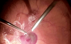
View
a narrated video of subretinal hemorrhage evacuation in RAMA.
Photo/video of RAMA Treatment courtesy of Alan Franklin, MD, PhD.
In 2009, Tsujikawa reported that RAMA can cause destruction of the foveal outer photoreceptor layer, resulting in poor visual outcome.1 This has led many surgeons to try and evacuate excessive subretinal hemorrhages, as seen in the accompanying video. This is a very tricky surgery that carries a high risk of complications, so it is reserved for cases where lack of intervention would almost certainly lead to poor visual outcome.
Notice how the surgeon is working in both the preretinal and subretinal space in this video. The surgeon starts by delaminating the posterior hyaloid with a forceps and the vitrectomy instrument. He then peels the internal limiting membrane and injects tissue plasminogen activator subretinally to liquefy the clot (nicely displayed on video). This procedure will conclude with a partial air/fluid exchange.
This year, there have been several published case reports on the use of anti-VEGF injections to speed the recovery of macroaneurysms, but this treatment is still considered new and unproven.2 At present, surgical intervention remains the prevailing approach.
There are several treatment options for RAMA if the macula is involved. If you come across this type of hemorrhage in your patient population, proper counseling and appropriate surgical consultation will often result in a high rate of visual recovery.
1. Tsujikawa A, Sakamoto A, Ota M, et al. Retinal structural changes associated with retinal arterial macroaneurysm examined with optical coherence tomography. Retina. 2009 Jun;29(6):782-92.
2. Cho HJ, Rhee TK, Kim HS et al. Intravitreal bevacizumab for symptomatic retinal arterial macroaneurysm. AJO. 2013 May;155(5):898-904.
