 |
History
A 27-year-old Caucasian female reported to the office with a chief complaint of “needing new glasses.” She had no other ocular or systemic issues, took no medications, was in good health and denied allergies of any kind.
Diagnostic Data
Her best-corrected entering visual acuities were 20/20 OD and 20/25 OS at distance and near. External examination was normal and no evidence of afferent pupillary defect was seen. Refraction corrected the issue, yielding improved vision at distance of 20/20 in both eyes.
Biomicroscopy of the anterior segment uncovered unusual ring-shaped opacities in both peripheral corneas (the pertinent clinical findings are demonstrated in photograph). Goldmann applanation tonometry measured 15mm Hg OU. Peripheral pathologies were seen OU.
Discussion
Additional studies might include gonioscopy and iris inspection to rule out angle abnormalities or maldeveloped iris tissue. New technologies using ultrasound biomicroscopy may also be helpful.
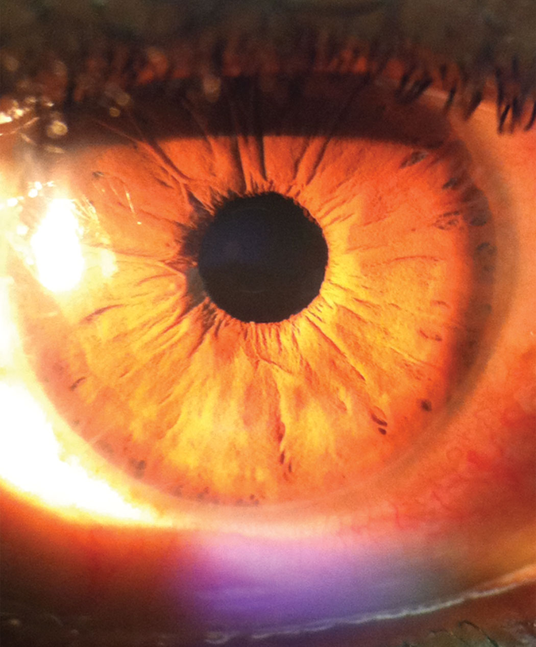 |
| This photo of the patient’s anterior presentation shows her unusual ring-shaped opacities in the peripheral cornea. Click image to enlarge. |
The diagnosis in this issue is posterior embryotoxin without evidence of iris mal-development or angle dysgenesis. Posterior embryotoxin is a thickened or hypertrophied Schwalbe’s ring (transition zone between the cornea and trabecular meshwork) that is anteriorly displaced and visible through the clear cornea as a sharply defined, concentric, white, ring opacity anterior to and inside the limbus.1-3 It is found in approximately 15% to 24% of normal eyes but may occur as a component of the anterior iris/angle developmental pathologies such as Axenfeld-Rieger’s anomaly.3 The term toxon is derived from the greek meaning “bow-like.” Alone it has no clinical or functional significance.
When associated with the irido-angular pathologies (Axenfeld’s anomaly, Reiger’s anomaly, Peter’s anomaly), multiple bridging peripheral iris strands and iris stromal hypoplasia can be identified.4-10 Interestingly, upwards of 10 hereditary disorders can be identified by characteristic iris, lens and corneal changes. Some of these include iris angle glaucoma disorders (Cogan Reese, Chandler syndrome and essential iris atrophy), Down’s syndrome (Brushfeld spots), Neurofibromatosis (Lisch nodules), chronic uveitis (Busacca/Koeppe nodules), heterochromia (Horner’s syndrome and Waaardenburg syndrome), diseases which produce leukocoria (retinoblastoma, persistent hyperplastic vitreous, retinal detachment, congenital cataract) and diseases which induce corneal changes such as Wilson’s disease (Kayser-Fleischer ring) and keratoconus (Fleisher ring).10
|
