Systemic hypertension (HTN) is a common health problem—affecting more than 800 million people worldwide—that often remains asymptomatic until late in the disease course.1 It is a major risk factor for both coronary artery disease and cerebrovascular accident.
|
|
|
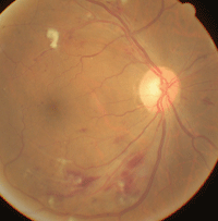
A patient with significant hypotensive retinopathy shows cotton-wool spots, striated retinal hemorrhages and narrowed retinal arterioles.
|
Although HTN has both genetic and environmental factors, the exact mechanism in the majority of affected individuals is largely unknown. Cardiac hypertrophy, heart failure, aortic dissection and renal failure are all systemic sequelae of the disease process.
In the eye, hypertensive ocular changes can be the initial finding in a patient with undiagnosed HTN, and may have sight-threatening consequences. In this article, we’ll review the ocular implications of hypertension.
Autoregulation in Retinal Vessels
Blood flow in the retina—as well as in the kidneys, brain, heart and skeletal muscle—is controlled by autoregulation, which is the intrinsic ability to maintain a constant blood flow despite changes in perfusion pressure. Thanks to autoregulation, retinal and other blood vessels constrict or dilate depending on hyper- or hypoperfusion status.4,5
Autoregulation operates within a certain range of perfusion pressure, and can be disrupted by alterations in blood pressure due to a variety of local and systemic causes. An increase or decrease in perfusion pressure beyond a critical autoregulatory range causes a breakdown of autoregulation. That is, autoregulation does not protect retinal vessels all of the time. The perfusion pressure may go above (malignant hypertension) or fall below (arterial hypotension) the critical range and thus subject the retinal tissue to ischemic damage.4,5
| Blood Pressure Norms4 | ||
| Normal |
Systolic: less than 120mm Hg Diastolic: less than 80mm Hg |
|
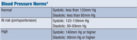 At risk (prehypertension) At risk (prehypertension)
|
Systolic: 120-139mm Hg
Diastolic: 80-89mm Hg |
|
| High |
Systolic: 140mm Hg or higher
Diastolic: 90mm Hg or higher |
Types of Hypertension
Blood pressure varies throughout the population based on age, gender, body mass index and diet.4 (See “Blood Pressure Norms,” below.)
• Essential hypertension. About 95% of HTN occurs as this form. Essential hypertension does not cause short-term problems, and is often called benign hypertension.
• Secondary hypertension. A small amount of HTN patients (5%) have this form. It’s caused by underlying conditions, such as renal or adrenal disease.4,5
• Malignant hypertension. Some patients have a rapid rise in blood pressure that, if untreated, may lead to death within a year or two. Malignant hypertension, also called accelerated hypertension, is characterized by severely elevated blood pressure (systolic greater than 200mg Hg, diastolic greater than 120mm Hg), renal failure, retinal hemorrhages and exudates with or without optic nerve head swelling. Malignant hypertension may occur in previously normotensive patients, but is usually superimposed on benign or secondary hypertension.4
Posterior Segment in HTN
During the early, acute phase of malignant hypertension, terminal arterioles may show two types of change. Dilation of the arterioles may occur, along with arteriolar occlusion, which eventually results in the formation of inner retinal ischemic “cotton-wool” spots and focal retinal capillary obliteration.
|
Public Health Implications of Hypertension Systemic hypertension puts patients at risk for heart disease and stroke, which are leading causes of death in the United States. We need to know about this because:
|
Focal intraretinal periarteriolar transudates are caused by a severe rise of arterial blood pressure. The retinal vasculature cannot properly autoregulate, causing focal dilation of precapillary retinal arterioles. Breakdown of the blood-retinal barrier occurs, causing increased permeability of the dilated arterioles to plasmatic deposits that flow into the retinal tissue.
The pathogenesis of optic neuropathy in malignant HTN has been a highly controversial subject. The various explanations can be divided into four categories:
• Disc edema due to raised intracranial pressure.
• Disc edema similar to hypertensive encephalopathy.
• Disc edema as a part of hypertensive retinopathy.
• Disc edema that is ischemic in nature.
Studies have shown that hypertensive optic neuropathy is primarily ischemic in nature. Similarly, clinical patterns of optic disc edema and optic disc changes in malignant hypertension are similar to those associated with anterior ischemic optic neuropathy (AION).5,6
Treatment and Management
The primary treatment of hypertensive retinopathy is systemic control of the arterial blood pressure with blood pressure medication and lifestyle changes.
The visual prognosis is excellent in mild to moderate cases. However, permanent visual loss may occur when there is persistent foveal edema and/or lipid deposition.
Be aware that a rapid reduction of blood pressure in a patient with hypertensive optic neuropathy may pose a risk of worsening ischemic damage to the optic nerve. A precipitous reduction in blood pressure will reduce perfusion to the optic nerve and central nervous system as a consequence of their autoregulatory changes, resulting in infarction of the optic nerve head and, potentially, acute ischemic neurologic lesions of the central nervous system.
A thorough systemic evaluation should focus on the etiology of the hypertension. Secondary causes should be ruled out in selected patients.
The eye is unique in that it allows direct observation of hypertensive consequences of the microvasculature in vivo. These ocular changes are of value in the management of systemic complications due to hypertension, including diabetes, cardiovascular, cerebrovascular and other systemic vascular diseases. n
1. Go AS, Mozaffarian D, Roger VL, et al; American Heart Association Statistics Committee and Stroke Statistics Subcommittee. Circulation. 2014 Jan 21;129(3):e28-e292.
2. Egan BM, Zhao Y, Axon RN. US trends in prevalence, awareness, treatment, and control of hypertension, 1988-2008. JAMA. 2010 May 26;303(20):2043-50.
3. Centers for Disease Control and Prevention. Vital signs: awareness and treatment of uncontrolled hypertension among adults—United States, 2003-2010. MMWR. 2012;61(35);703-9.
4. Kumar V, Abbas A, Fausto N. Robbins and Cotran’s Pathologic Basis of Disease, 7th ed. Philadelphia: Elsevier Saunders; 2005.
5. Spencer WH. Systemic diseases with retinal involvement: vascular diseases. In: Ophthalmic Pathology: An Atlas and Textbook (CD-ROM), 4th ed. WB Saunders Co.; 1995.
6. Quillen DA, Blodi BA, Bennett TJ. Clinical Features of Retinal Disease. In: Quillen DA, Blodi BA (eds.). Clinical Retina. Chicago: AMA Press; 2002:136-7.
|
Retinal and Choroidal Lesions In Hypertension5,6 Retinal vascular lesions
Extravascular retinal lesions Retinal hemorrhages (usually situated in the nerve fiber layer and commonly in the distribution of the radial peripapillary retinal capillaries, although they could be located anywhere in the fundus)
RPE/Choroidal lesions
|
