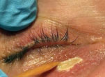
While some patients with xanthelasma ask their optometrist, “what are these yellow spots,” others may be completely unaware of the plaques that have formed. In either case, clinicians must educate patients on the systemic implications associated with these plaques.
Spotting the Spots
Xanthelasma, also known as xanthoma, are benign soft yellow-orange plaques. They vary in size and are often found on the eyelids but can present on any cutaneous surface. Frequently, both the upper and lower lids are involved.1 Onset of xanthelasma typically begins during the fourth or fifth decade of life and is more prevalent in women.1
Fifty percent of patients presenting with xanthelasma palpebrarum have hyperlipidemia.2 For patients younger than 40, additional testing is indicated to rule out potential lipoprotein and apolipoprotein abnormalities.2 Patients with xanthelasma are at a higher risk of atherosclerosis and should have a lipid panel completed so that their primary care provider can monitor for any underlying systemic disease.2
 |
| This patient chose to undergo chemical cauterization to resolve his xanthelasma. |
Therapies
Several options exist to remove xanthelasma. Methods include: traditional surgical excision, topical trichloroacetic or bichloracetic acid, liquid nitrogen, electrodessication, cauterization and several laser options.1,3,4
Treatment methods using chemicals or lasers can result with permanent hypopigmentation of the skin after treatment.3 This effect is an important consideration when recommending a treatment method to a patient. These treatment options are contraindicated in individuals with darker complexion. Traditional surgical excision is the treatment of choice for these patients to optimize cosmetic outcomes.3
Bichloracetic acid is one option for patients looking for simple, effective treatment. In a study of 25 xanthelasma plaques, 85% were resolved after initial treatment and 72% remained resolved over five years.1 In cases where recurrence was observed, the patients had associated hyperlipidemia.1
Case in Point
A 47-year-old Caucasian male presented with a single xanthelasma plaque on his lower right lid. The patient had a lipid panel revealing hyperlipidemia and elevated triglycerides. Bichloracetic acid treatment was performed to remove his lesion. The patient was given one drop of topical 0.5% proparacaine into his right lower cul-de-sac. His skin was cleaned using an alcohol wipe. Topical or local anesthetic is usually not indicated for this method of treatment. A generous amount of petroleum jelly, applied with a cotton-tip applicator, was used to protect the uninvolved healthy tissue surrounding the plaque. Bichloracetic acid was then applied onto the surface of the plaque using a wooden applicator. Once liquification and pallor of the target tissue is observed, the chemical cauterization is complete. The petroleum jelly is then removed with a clean cotton-tip applicator.
The patient was directed to not disturb the treated tissue. The lesion will shortly become necrotic and slough off.
Ms. Westall is a fourth-year student at Pacific University College of Optometry.
Dr. Skorin is a consultant in the Department of Surgery, Community Division of Ophthalmology in the Mayo Clinic Health System in Albert Lea, MN.
1. Haygood LJ, Bennett JD, Brodell RT. Treatment of xanthelasma palpebrarum with bichloracetic acid. Am Soc Dermatol Surg, 1998;24:1027-31. 2. Bergman R. The pathogenesis and clinical significance of xanthelasma palpebrarum. J Am Acad Dermatol, 1994;30:236-42. 3. Laftah Z, Al-Niaimi F. Xanthelasma: An update on treatment modalities. J Cutan Aesthet Surg. 2018;11:1-6. 4. Obradovic B. Surgical treatment as a first option of the lower eyelid xanthelasma. J Craniofacial Surg. 2017;28:678-9. |

