 A 60-year-old white female presented with symptoms of blurred vision (OD > OS) and bilateral floaters that had persisted for the last six months.
She also noted increased difficulty seeing at night. Her systemic history was significant for hypertension and high cholesterol, for which she was properly medicated.
On examination, her best-corrected visual acuity measured 20/100 OD and 20/30 OS. Confrontation visual fields were full to careful finger counting OU. Her pupils were equally round and reactive, with no evidence of afferent defect.
The anterior segment evaluation was significant for early nuclear sclerotic and trace posterior subcapsular cataracts OU.
Dilated fundus exam showed a significant vitritis in both eyes. The optic nerves appeared healthy, with small cups and good rim coloration and perfusion OU. The arteries and veins were slightly attenuated. There was no foveal light reflex in either macula. Additionally, we detected a mild epiretinal membrane OU.
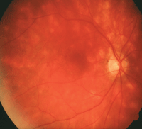
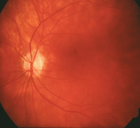
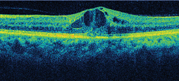
On indirect ophthalmoscopy, we noted obvious retinal changes (figures 1 and 2). Further, we obtained a spectral domain optical coherence tomography (SD-OCT) scan (figure 3).
Take the Retina Quiz
1. What does the SD-OCT scan reveal?
a. Neurosensory retinal detachment.
b. Cystoid macular edema (CME).
c. Retinoshisis.
d. Stage 1 macular hole.
2. At which retinal level are the depigmented lesions located?


1, 2. Posterior pole and midperiphery of both eyes exhibit hazy media, vessel attenuation and hypopigmented spots (OD left, OS right).
a. Choroid.
b. Retinal pigment epithelium (RPE).
c. Sensory retina.
d. Both sensory retina and RPE.
3. What is the likely diagnosis?
a. Multifocal choroiditis and panuveitis.
b. Serpiginous choroiditis.
c. Vitiliginous chorioretinitis.
d. Syphilis.
4. What additional tests would help confirm the diagnosis?
a. Fluorescein angiography.
b. Blood testing for HLA-A29.
c. Blood testing for HLA-B9.
d. Angiotensin-converting enzyme (ACE).
5. What is the best treatment option?
a. Corticosteroids.
b. Observation.
c. Immunosuppressive agents.
d. Both a and c.
For answers, see below.
Discussion
We diagnosed our patient with vitiliginous chorioretinitis, a rare inflammatory condition of the choroid and retina. The condition originally was termed “birdshot retinochoroidopathy” in 1980, because the scattered displacement of the associated lesions was reminiscent of a shotgun blast.1
Meanwhile, around the same time, J. Donald M. Gass, MD, of the Bascom Palmer Eye Institute in Miami, used the term vitiliginous chorioretinitis to describe the condition because he believed the depigmented retinal lesions resembled skin lesions observed on patients with vitilligo.2 Since then, both terms have been used interchangeably by academics and practicing clinicians.
In the early reports, vitiliginous chorioretinitis was thought to occur predominantly in women.1 Today, the condition is understood to occur in both men and women in the fifth to seventh decade of life.2 The most common symptoms include blurred vision, increased floater volume and photopsia.2 In advanced disease progression, patients frequently report night blindness and color vision loss.2

3. An SD-OCT scan of the right eye. Can you discern any macular changes?
The hallmark of vitiliginous chorioretinitis is significant vitritis (accounting for the increased floaters) and multifocal patches of depigmented or hypopigmented lesions that may be creamy yellow or orange in color. The ill-defined patches typically are round or oval in shape. Some will appear elongated in a pattern that radiates toward the peripheral fundus.
The legions’ striking feature is the lack of chorioretinal scaring or hyperpigmentation at the margins, which often are seen in other inflammatory conditions. The disease originates in the choroid and later involves the RPE. Interestingly, there does not appear to be any thinning within the RPE or choroid in the depigmented areas.2
The diagnosis of vitiliginous chorioretinitis usually is made based on the clinical presentation; however, there also is a pronounced association with the HLA-A29 antigen. More specifically, at least 90% of patients with vitiliginous chorioretinitis test positive for HLA-A29 upon examination––suggesting an autoimmune mechanism as well as a genetic predisposition.2 In fact, this association is so strong that you should consider a diagnosis of saroidosis or another granulomatous condition if the patient tests negative for the HLA-A29 antigen.
Vitiliginous chorioretinitis is a chronic, slowly progressive condition that exhibits periods of remission and exacerbation. Typically, patients lose vision from cystoid macular edema (as we documented in our patient), which results from chronic inflammation. As a consequence, management is aimed at quieting the inflammation.
Corticosteroids have been the mainstay treatment option, but have yielded limited success. Patients may note visual improvement as a result of CME resolution, but will not exhibit a decrease in lesion number or severity. Immunosuppressive agents, such as methotrexate, mycophenolate mofetil and cyclosporine, also have been used alone or in combination with corticosteroids for long-term treatment.2
We treated our patient with pulsed, high-dose oral steroids and low-dose methotrexate. Her CME resolved and her vision returned to 20/25 OU. However, during the ensuing years, she continued to experience recurrences and exacerbations while on immunosuppressive agents.
1. Ryan SJ, Maumenee AE. Birdshot retinochoroidopathy. Am J Ophthalmol. 1980 Jan;89(1):31-45.
2. Agarwal A. Inflammatory Disease of the Retina. In: Gass’ Atlas of Macular Diseases. 5th ed. Elsevier Saunders: Philadelphia; 2012:1038-43.
Answers
1. b
2. a
3. c
4. b
5. d

