 |
When floaters begin to interfere with patients’ work, they are going to begin looking for any solution. Such was the recent case of a physician—a friend of a friend—who felt his floaters were beginning to interfere with his ability to practice within his specialty. He had consulted an ophthalmologist who promoted a laser treatment designed to rid patients of floaters altogether. The patient was hesitant and contacted me. Although floaters are a common nuisance, their causes and treatment options are poorly understood by patients—even some who are physicians themselves.
This column provides a quick breakdown of what to tell patients experiencing this often worrying, but rarely dangerous, issue.
What are Floaters?
The vitreous is a hydrated and acellular gel constituting the bulk of the globe contents. It consists of 98% water with the remainder collagen and hyaluronan forming a clear gel. At birth, the vitreous gel is quite firm, but with aging (and myopic vitreopathy), this firm gel liquefies, forming pockets of liquid vitreous known as lacunae. This liquefaction occurs from the dissociation of hyaluronan from the collagen fibrils forming the supportive network of the vitreous gel. This allows for the aggregation of the smaller collagen fibrils into macroscopic fibers, which subsequently diffract light. The accumulation of lacunae transforms the vitreous gel into a liquid consistency. This facilitates collapse of the vitreous and the development of a posterior vitreous detachment (PVD), occurring as a separation of the posterior vitreous cortex from the internal limiting membrane of the retina. This process begins posteriorly and progresses anteriorly to the vitreous base. Additionally, lacunae increase vitreous heterogeneity and scattering of light at the gel-liquid interface.1
 |
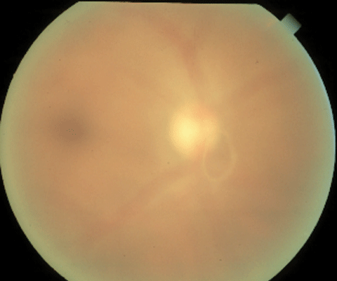
| Weiss ring in an acute PVD causing a complaint of floaters. Click image to enlarge. |
Floaters, or symptomatic vitreous opacifications (SVO), are packed bundles of collagen fibrils that first appear in the central vitreous where they often have a linear configuration, becoming more numerous, thickened and irregular with increasing age and axial myopia.1 The walls of the liquefied lacunae interfere with photon transmission to the retina, contributing to the common symptom of floating spots. They are more visible when viewed against a bright source such as a sunny sky or a white background. They are even more noticeable when situated in the visual axis. The onset of PVD increases the scatter of light and an increase in symptoms as more collagen aggregates are directed toward the visual axis. Many patients undergoing PVD ultimately adapt to the SVOs, most likely due to the fact that the Weiss ring eventually settles inferiorly and anteriorly, affecting the penumbra cast upon the retina in a less symptomatic form.2
Get Used to It?
SVOs are among the most common, and most dismissed, complaints you’ll encounter. Most practitioners, upon confirming there is no tractional retinal pathology, explain to the patient that adaptation will occur and there is nothing to worry about. In most cases, patients either adapt or accept and pursue the issue no further. Indeed, as the opacifications move out of the visual axis or locate more anteriorly with forward rotation of the collapsed vitreous, the symptoms will greatly decrease, usually to an acceptable level. However, there is a small subset of patients who do not adapt and have a greatly reduced quality of life from SVOs despite excellent Snellen acuity. This makes determination of the most effective and appropriate treatments difficult to scientifically assess. Thus, virtually all interventions are patient driven.
However, decreased quality of life comes from reduced contrast sensitivity, decreased driving ability and impaired facial recognition.1 Up to two thirds of patients with SVOs have moderate to extreme difficulty in reading small print or driving at night.3 In patients younger than 55 years old, these effects are so deleterious that they were willing to undergo a procedure that carried a risk of 7% blindness to be cured of SVOs.4
Currently, two non-observational management options for SVOs exist. These are Nd:YAG laser vitreolysis and vitrectomy.
Vitreolysis
The least invasive option to combat floaters is laser vitreolysis, but the procedure may have complications and limitations. Nd:YAG vitreolysis is performed by focusing the laser onto vitreal opacities visible at the biomicroscope. The aim is to reduce the volume of the individual floaters by disintegrating them down to smaller fragments, by cutting longer strands into shorter strands or by cutting strands that suspend larger floaters, allowing them to dislodge from the visual axis.2 Nothing is actually removed from the vitreous. The large floaters are reduced to smaller floaters or laser induced incisions in collagen strands may allow large opacities to move elsewhere. There is no guarantee that the patient won’t still have SVOs after the procedure. Indeed, they may just have different floaters that are hopefully not as bothersome. Also, due to the risk of laser-induced damage, only SVOs greater than 4mm away from the retina and lens are typically treated. Hence, Nd:YAG laser vitreolysis may not be able to treat the most symptomatic floaters.2 It is difficult to gauge outcomes from this procedure, but subjective success rate of laser vitreolysis appears to be relatively low.1,5
 |
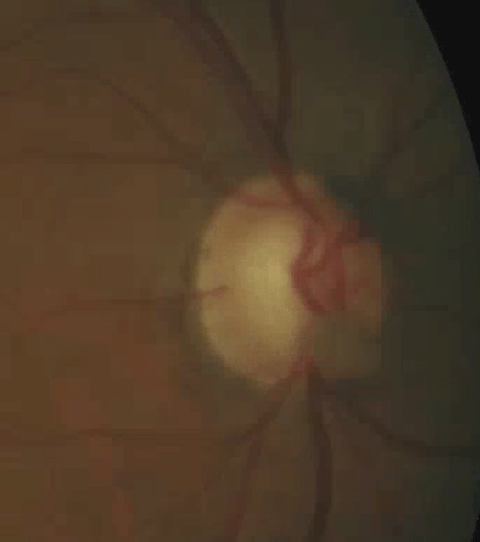
| Symptomatic posterior pole floater. |
While laser vitreolysis is the lesser invasive option, it is not without risk. Complications of Nd:YAG laser treatment for other conditions include induction of retinal breaks and detachment as well as cystoid macular edema. One study reported on a refractory glaucoma occurring after laser vitreolysis for SVOs requiring surgical intervention to lower IOP.6
Vitrectomy
Pars plana vitrectomy (PPV) is a more invasive, but possibly more effective option.2 Smaller gauge instruments have allowed for self-sealing sutureless sclerotomies with a faster recovery and reduced rate of complications.2
Sutureless 25-gauge pars plana vitrectomy for symptomatic vitreous floaters improves visual acuity, resulting in a high patient satisfaction quality-of-life survey, with a low rate of postoperative complications.1 Sutureless pars plana vitrectomy should be considered as a viable means of managing patients with symptomatic vitreous floaters. Additionally, because the SVOs are actually being removed, PPV reported success rates are very high.7-9 In one series, the patient-reported success rate was 85%. Tellingly, 87% of patients said that they would recommend PPV to friends with similar complaints.3
Of course, PPV carries a greater risk of serious complications, such as induction of retinal tears and detachment, choroidal hemorrhage, proliferative vitreoretinopathy, endophthalmitis and, most commonly, cataracts, occurring in 50% to 75% of cases postoperatively.1,10 Retinal tears and detachment are often associated with simultaneous induction of PVD during PPV. Use of small gauge instruments and not inducing PVD during the procedure greatly reduces these risks.
While Nd:YAG vitreolysis is a less invasive option, it is not without risks and the success rate is suspect. The use of smaller gauge instrumentation and sutureless PPV, combined with a highly successful, definitive outcome, appears to have lowered the threshold for performing this procedure on patients with SVOs who have reduced quality of life and function. Ultimately, it rests with the patient to decide if the benefits of an invasive surgery outweigh the risks.
|
1. Milston R, Madigan MC, Sebag J. Vitreous floaters: Etiology, diagnostics, and management. Surv Ophthalmol. 2016;61(2):211-27. 2. Ivanova T, Jalil A, Antoniou Y, et al. Vitrectomy for primary symptomatic vitreous opacities: an evidence-based review. Eye (Lond). 2016 May;30(5):645-55. 3. de Nie KF, Crama N, Tilanus M, et al. Pars plana vitrectomy for disturbing primary vitreous floaters: clinical outcome and patient satisfaction. Graefes Arch Clin Exp Ophthalmol. 2013;251(5):1373-82. 4. Wagle A, Lim W, Yap T, et al. Utility values associated with vitreous floaters. Am J Ophthalmol. 2011;152(1):60-5. 5. Delaney Y, Oyinloye A, Benjamin L. Nd:YAG vitreolysis and pars plana vitrectomy: surgical treatment for vitreous floaters. Eye (Lond). 2002;16(1):21-6. 6. Cowan L, Khine K, Chopra V, et al. Refractory open-angle glaucoma after neodymium-yttrium-aluminum-garnet laser lysis of vitreous floaters. Am J Ophthalmol. 2015;159(1):138-43. 7. Mason JO 3rd, Neimkin MG, Mason JO 4th, et al. Safety, efficacy, and quality of life following sutureless vitrectomy for symptomatic vitreous floaters. Retina. 2014;34(6):1055-61. 8. Sommerville DN. Vitrectomy for vitreous floaters: analysis of the benefits and risks. Curr Opin Ophthalmol. 2015;26(3):173-6. 9. Sebag J, Yee K, Wa C, et al. Vitrectomy for floaters: prospective efficacy analyses and retrospective safety profile. Retina. 2014;34(6):1062-8. 10. Henry CR, Schwartz SG, Flynn HW Jr. Endophthalmitis following pars plana vitrectomy for vitreous floaters. Clin Ophthalmol. 2014;8:1649-53. |

