 |
A 66-year-old woman presented complaining of a large, painless lump in her left upper lid that developed rather rapidly over several days beginning two months earlier. Though the lesion was painless now, she did experience some moderate pain while it was developing. She had been putting hot water on it to no avail.
Evaluation
Her best-corrected visual acuity was 20/20 in each eye. The remainder of her examination was normal, except for biomicroscopically visible crusting about the eyelashes, blocked meibomian glands and a grossly visible, painless, hard, focal lump in her left upper eyelid. Based upon appearance and history, it was clear that she had developed a chalazion, most likely secondary to chronic blepharitis. There are several options for treating this patient and virtually every doctor has a “favored” approach to managing chalazia. However, does the science support any or any of these “can’t-miss” treatments?
Conservative Therapies
Chalazion is a common inflammatory condition of the eyelid. Two patterns of granulomatous inflammation represent the spectrum of changes in its clinical course. Mixed-cell granulomas consisting of neutrophils, lymphocytes, plasma cells, macrophages, giant cells and granulation tissue may be present.1,2 Additionally, you may see suppurating granulomas characterized by epithelioid cell granulomas with numerous neutrophils in a proteinaceous background.1,2 The cells involved in these lesions are steroid sensitive.1,2
Chalazia (plural of chalazion) are typically caused by blockage of the meibomian glands and chronic lipogranulomatous inflammation. It can affect patients of any age, race or gender. Common complaints are poor cosmesis, local irritation and, in cases of large lesions, mechanical ptosis and corneal astigmatism.3
Conservative management can include lid scrubs (either with baby shampoo or commercially prepared scrubs), hot compresses with or without digital massage, topical antibiotic solutions and ointments, oral antibiotics or a combination of antibiotic or steroid solutions and ointments.4-6 However, the effects of these approaches aren’t always clear. One study shows simple lid hygiene resulted in clinical chalazia cure in 80% of cases.6 Another shows hot compresses and lid hygiene together had a 43% cure rate.5 In a 2006 investigation hot compresses, lid hygiene and antibiotic ointment QID, shows a 58% cure rate.4
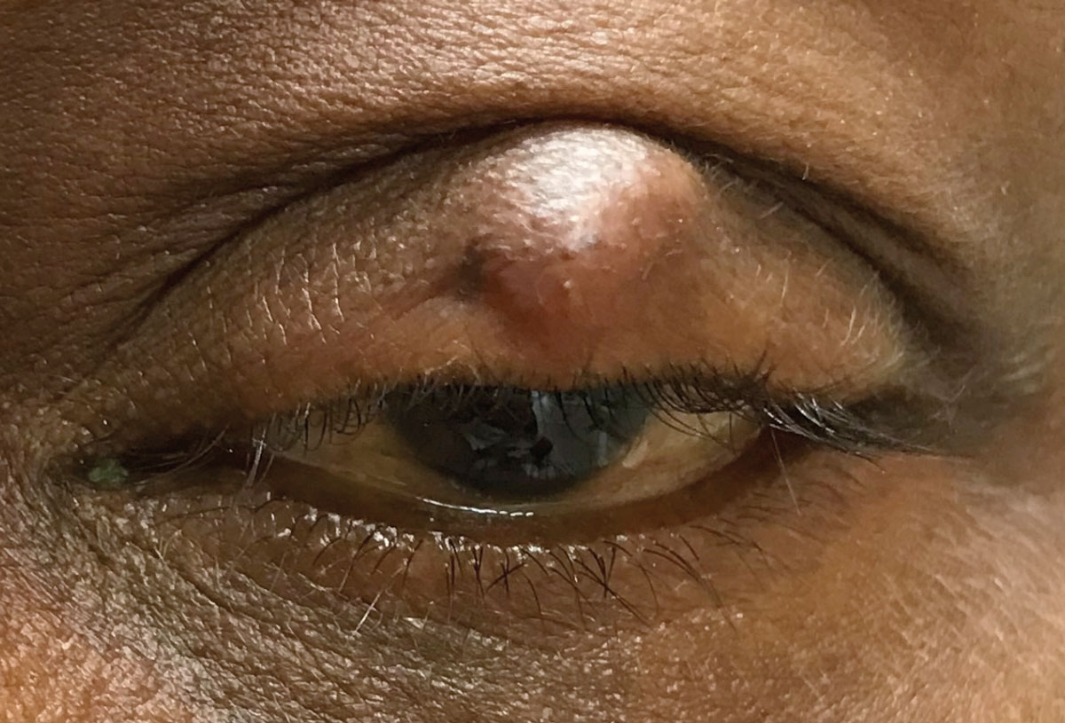 |
| A lid lump like this patient’s may resolve with conservative therapy, but if it doesn’t, steroids and surgery are options. Click image to enlarge. |
More recent research looked at 149 patients with one or more chalazia on separate eyelids and randomized them to receive therapy involving hot compresses only, hot compresses plus tobramycin drops and ointments, and hot compresses plus tobramycin/dexamethasone drops and ointment over four to six weeks. Treated with hot compress alone, 21% saw complete lesion resolution. Adding tobramycin drops and ointments to hot compresses worked out for 16% of subjects and hot compresses plus tobramycin/dexamethasone drops and ointments helped 18% achieve resolution.7
None of these differences in treatment could be considered significant. Lesions that completely resolved had a statistically significant lower pretreatment duration of 1.5 months compared with lesions that did not completely resolve with lesions present over 2.2 months. This report shows that hot compresses alone or in combination with tobramycin or tobramycin/dexamethasone drops and ointment are all effective first-line treatments; however, lesions present longer than two months are less likely to resolve with these conservative therapies alone.7
While “conservative” therapies are non-invasive and exceedingly safe, they clearly do not work for every patient.
Steroids
The inflammatory cells that comprise chalazia are steroid sensitive, which is why some research is considering intralesional steroid injection as a management option.2-11 Intralesional injection involves the injection of 0.1ml to 0.3ml of triamcinolone actetonide (5mg/ml to 40mg/ml) from a conjunctival approach.3,4,8 Like conservative therapies, no clear delineations as to the optimal amount and concentration of steroid exist. However, the success rates for this management modality is typically higher than for conservative therapy.4-9 A 2006 study saw a 94% cure rate (vs. 58% for conservative therapy) with intralesional injection of triamcinolone.4 Another investigations achieved an 80% success rate after two injections.9
While intralesional injection of triamcinolone is generally safe, significant complications can occur. Skin depigmentation is a common occurrence following intralesional injection in dark-skinned patients.4,9,11 Also, inadvertent globe perforation is a possibility.12 Rarely, microembolization by steroid particles can result in retinal and choroidal infarction with subsequent permanent vision loss.12
Surgery
Incision and curettage remains an option. The lesion can be surgically removed, typically through a palpebral conjunctival approach, with the use of scalpel and chalazion clamp following an injection of anesthetic. Thermal cautery is often performed immediately following surgical excision, but this is typically the surgeon’s choice, as it does not appear to reduce recurrence rate.13 Cure rates with surgical excision are between 90% and 100%, though more than one surgery may be necessary.8,9 Surgical excision is typically the recommended procedure for lesions that are larger than 11mm and chronic (lasting more than eight months).2
While highly successful, surgical excision also has potential complications. If excision goes through the dermis, scarring is possible. Further, inadvertent globe perforation may occur during chalazion excision.14 Surgical excision remains an option if conservative or intralesional injection fail to resolve the condition.
Comparing intralesional steroid injection to surgical curettage in a meta-analysis, researchers found that for a single procedure, surgical curettage was more successful than steroid injection at achieving resolution.15 If multiple procedures were necessary, then the difference in success between steroid injection and surgical curettage was reduced.15
Intralesional steroid injection and surgical curettage shows similar results.16 A single triamcinolone acetonide injection followed by lid massage is almost as effective as incision and curettage.17 A study of chronic chalazia that were unresponsive to medical treatment, noted that lesions responded well to both steroid injection and surgical curettage.
In our case, conservative therapy with hot compresses alone was recommended. Due to her skin pigmentation, steroid injection was not advocated. Ultimately, conservative therapy did not result in any significant improvement and she underwent successful surgical excision.
1. Dhaliwal U, Arora VK, Singh N, et al. Cytopathology of chalazia. Diagn Cytopathol. 2004;31(2):118-22. 2. Dhaliwal U, Bhatia A. A rationale for therapeutic decision-making in chalazia. Orbit. 2005;24(4):227-30. 3. Ben Simon GJ, Huang L, Nakra T, et al. Intralesional triamcinolone acetonide injection for primary and recurrent chalazia: is it really effective? Ophthalmology. 2005;112(5):913-7. 4. Chung CF, Lai JS, Li PS. Subcutaneous extralesional triamcinolone acetonide injection versus conservative management in the treatment of chalazion. Hong Kong Med J. 2006;12(4):278-81. 5. Garrett GW, Gillespie ME, Mannix BC. Adrenocorticosteroid injection vs. conservative therapy in the treatment of chalazia. Ann Ophthalmol. 1988;20(5):196-8. 6. Perry HD, Serniuk RA. Conservative treatment of chalazia. Ophthalmology. 1980;87(3):218-21. 7. Wu AY, Gervasio KA, Gergoudis KN, et al. Conservative therapy for chalazia: is it really effective? Acta Ophthalmol. 2018;96(4):e503-e509. 8. Mustafa TA, Oriafage IH. Three methods of treatment of chalazia in children. Saudi Med J. 2001;22(11):968-72. 9. Ahmad S, Baig MA, Khan MA, et al. Intralesional corticosteroid injection vs surgical treatment of chalazia in pigmented patients. J Coll Physicians Surg Pak. 2006;16(1):42-4. 10. Kaimbo Wa Kaimbo D, Nkidiaka MC. Intralesional corticosteroid injection in the treatment of chalazion. J Fr Ophtalmol. 2004;27(2):149-53. 11. Ho SY, Lai JS. Subcutaneous steroid injection as treatment for chalazion: prospective case series. Hong Kong Med J. 2002;8(1):18-20. 12. Thomas EL, Laborde RP. Retinal and choroidal vascular occlusion following intralesional corticosteroid injection of a chalazion. Ophthalmology. 1986;93(3):405-7. 13. Sendrowski DP, Maher JF. Thermal cautery after chalazion surgery and its effect on recurrence rates. Optom Vis Sci. 2000;77(11):605-7. 14. Shiramizu KM, Kreiger AE, McCannel CA. Severe visual loss caused by ocular perforation during chalazion removal. Am J Ophthalmol. 2004;137(1):204-5. 15. Aycinena AR, Achiron A, Paul M, Burgansky-Eliash Z. Incision and Curettage Versus Steroid Injection for the Treatment of Chalazia: A Meta-Analysis. Ophthalmic Plast Reconstr Surg. 2016;32(3):220-4. 16. Singhania R, Sharma N, Vashisht S, Dewan T. Intralesional Triamcinolone Acetonide (TA) Versus Incision and Curettage (I & C) for Medium and Large Size Chalazia. Nepal J Ophthalmol. 2018;10(19):3-10. 17. Goawalla A, Lee V. A prospective randomized treatment study comparing three treatment options for chalazia: triamcinolone acetonide injections, incision and curettage and treatment with hot compresses. Clin Exp Ophthalmol. 2007;35(8):706-12. 18. Nabie R, Soleimani H, Nikniaz L, et al. A prospective randomized study comparing incision and curettage with injection of triamcinolone acetonide for chronic chalazia. J Curr Ophthalmol. 2019;31(3):323-6. |
