 Blood clotting is a natural and necessary process in the body—we rely on it to stop us from bleeding too much when an injury occurs. The wound triggers the clotting process, drawing certain chemicals in the blood to the site, where they solidify into a clot and plug the injured part of the blood vessel. Another set of natural chemicals in the blood counterbalance these clotting factors to stop the blood from clotting too much.
When too much clotting factor upsets this natural balance, it’s known as thrombophilia (or a hypercoagulable state).1 Many patients who have this condition never experience any medical issues as a result—thrombophilia simply increases the likelihood that they could develop an unwanted blood clot. When we encounter thrombotic conditions in the eye, such as non-arteritic anterior ischemic optic neuropathy (NAION), retinal artery occlusion and retinal vein occlusion, we must consider the possibility of thrombophilia.
What Causes Thrombophilia?
Hypercoagulable states may be inherited or acquired. Acquired hypercoagulable states may occur as a result of surgery, trauma, medications, pregnancy or medical conditions, such as cancer, diabetes and obesity.1,2
As optometrists, we need to be particularly aware of antiphospholipid antibody syndrome, an acquired, hypercoagulable autoimmune disorder that can not only lead to retinal vascular occlusion, but also life-threatening complications, such as heart attack and stroke.2
Hereditary Hypercoagulable Conditions1-4
• Factor V Leiden gene mutation
 Kevin A. Somerville © US Pharmacist 2003
• Prothrombin gene mutation
• Elevated homocysteine
•
Abnormal fibrinolytic system, including hypoplasminogenemia,
dysplasminogenemia and elevated plasminogen activator inhibitor (PAI-1)
• Deficiencies of proteins that prevent clotting (antithrombin, protein C and protein S)
• Elevated fibrinogen or dysfunctional fibrinogen (dysfibrinogenemia)
• Elevated factor VIII and other factors, including IX and XI
With inherited forms of thrombophilia (see “Hereditary Hypercoagulable Conditions,” below), patients are genetically predisposed with a tendency to form unwanted clots. The most common hereditary blood clotting disorder in the United States, Factor V Leiden gene mutation, affects 4% to 10% of whites.3,4 Genetically at-risk patients who develop blood clots usually do so when additional risk factors are present, such as obesity, smoking, oral contraceptives and hormone replacement therapy.1,3,4
Diagnostic Work-up
As always in cases of suspected systemic disease, perform a careful evaluation of the patient’s personal and family medical histories. A targeted review of systems for hypercoagulable states should include:1,3,4
- Family history of abnormal blood clotting.
- Abnormal blood clotting in patients younger than age 50.
- Thrombosis in unusual sites, such as veins in arms, liver, intestines, kidney and brain.
- History of “idiopathic” blood clots.
- Recurrent blood clots.
- Multiple miscarriages.
- Stroke at a young age.
- Myocardial infarction.
Although no single test can diagnose thrombophilia, we can perform a battery of lab tests to rule out or gather more information about the condition. (See “Ruling Out Thrombophilia.”)
To help diagnose inherited hypercoagulable states, we can order genetic tests for Factor V Leiden (activated protein C resistance) and prothrombin gene mutation (G20210A), as well as testing antithrombin activity, protein C activity, protein S activity and fasting plasma homocysteine levels.5-8
To help diagnose acquired hypercoagulable states, we can order anticardiolipin antibodies (ACA) or beta-2 glycoproteins and lupus anticoagulants (LA). Heparin antibody testing should be performed in patients who develop low platelet counts while exposed to heparin.2,5,6
The most appropriate lab tests to exclude a diagnosis of thrombophilia include:2,4
Ruling Out Thrombophilia
The Eye in Thrombophilia
The prevalence of inherited hypercoaguable states is much higher in patients with venous thromboembolism (which includes CRVO) as compared to the general population.9 For instance, the 5% incidence of Factor V Leiden in the general population jumps to 20% to 50% in patients with venous thromboembolism, while prothrombin G20210A jumps from 2% to 3% up to 6% to 8%.9
The prevalence of hyperhomocysteinemia is 5% to 10% in the general population, but doubles to 10% to 20% in those with venous thrombotic disease.9 Coupled with acquired hypercoagulation, the prevalence numbers become even more significant.
Hypercoagulable states may give rise to local thrombotic lesions in discrete segments of veins or arteries; therefore, focal thrombosis may involve the vessels that supply the retina, optic nerve, or both. It is important for the optometrist to be aware of the major inherited genetic mutations that lead to thrombophilia, especially in cases of young patients with diagnosed central retinal vein occlusion lacking any positive systemic history.
Retinal vein occlusion has been reported in association with Factor V Leiden, hyperhomocysteinemia, protein C deficiency, antithrombin deficiency, protein S deficiency and antiphospholipid antibody syndrome.9 Retinal arterial occlusion has been linked with hyperhomocysteinemia and elevated lipoprotein(s), while NAION has been reported in patients with Factor V Leiden and elevated lipoprotein(s).9
Standard treatment and management protocols for RVO/RAO/NAION apply. In addition, appropriate medical treatment for the systemic cause is necessary and may help prevent recurrences of thromboembolic disease in the eye and elsewhere.
Treatment Approaches
Medical treatment is indicated when a blood clot develops in a vein or artery (including retinal vein and retinal arterial occlusion).5-7 Anticoagulant medications are used to prevent systemic problems, such as heart attack or stroke, that might occur at a later time.
The most commonly used drugs include:
- Aspirin, which has an anti-platelet effect by inhibiting the enzyme cyclooxygenase, which in turn impedes thromboxane A2, an important intermediary involved in the clotting process.
- Coumadin (warfarin, Bristol-Myers Squibb), which comes in tablet form.
- Heparin, a liquid medication delivered by either intravenous or subcutaneous injections.10 (Low-molecular weight heparin is injected subcutaneously once or twice a day and can be taken at home.)
- Arixtra (fondaparinux, GlaxoSmithKline), which is injected subcutaneously.
In taking the proper steps toward screening and detection, we can help the patient and potentially affected family members get appropriate treatment and decrease their chances of life-threatening vascular disease, such as pulmonary embolism, myocardial infarction and deep vein thrombosis.
•
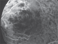
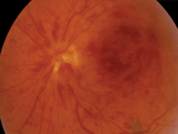
Diagnostic data. Best-corrected visual acuity was 20/20 OD and 20/50
OS. Dilated funduscopy revealed a non-ischemic central retinal vein
occlusion with mild foveal edema OS (figures 1 and 2).
•
Management. We scheduled the patient to return in two weeks for
follow-up of the CRVO. In the meantime, she discontinued the birth
control med after consultation with her primary care physician and
gynecologist. Because the patient recently had a complete physical
examination showing no diabetes, hypertension or hyperlipidemia, we
ordered a targeted blood work-up to rule out hypercoaguable states.
Results
of these tests were positive for the Factor V Leiden gene mutation. We
referred her to a hematologist, who prescribed aspirin therapy as the
initial medical management. The patient’s immediate family members were
evaluated (and possibly treated) for any genetic blood clotting
mutations.
At her two-month follow-up visit, the macular edema had
reduced (figure 3) and visual acuity improved to 20/30. She will return
for follow-up in a month.
Case Report: Sudden Vision Loss Suggests Hypercoagulable State
• History. A healthy 36-year-old white
female presented with a chief complaint of sudden vision loss in her
left eye, which occurred earlier that morning. Her medications included
an oral contraceptive, and she reported a family history of “blood
clots” and heart disease. Her personal health history was negative for
hypertension, diabetes, dyslipidemia and heart disease.


1,2. Dilated funduscopy revealed that our patient had a non-ischemic central retinal vein occlusion with mild foveal edema OS.

3. OCT images showed the macular edema had reduced at her two-month follow-up.
1. Cotran RS, Mitchell RN. Hemodynamic disorders, thrombosis and shock. In: Cotran RS, Kumar V, Collins T. Pathologic Basis of Disease. 6th ed. Philadelphia: W.B. Saunders Company; 1999.
2. Durrani OM, Gordon C, Murray PI. Primary anti-phospholipid syndrome (APS): current concepts. Surv Ophthalmol. 2002 May-Jun;47(3):215-38.
3. Bonner H, Bagg A, Cossman J. The blood and the lymphoid organs. In: Rubin E, Farber JL (eds). Pathology. 3rd ed. Philadelphia: Lippincott Williams & Wilkins; 1998:1050-151.
4. Price DT, Ridker PM. Factor V Leiden mutation and the risks for thromboembolic disease: a clinical perspective. Ann Intern Med. 1997 Nov;127(10):895-903.
5. Gottlieb JL, Blice JP, Mestichelli B, et al. Activated protein C resistance, factor V Leiden, and central retinal vein occlusion in young adults. Arch Ophthalmol. 1998 May;116(5):577-9.
6. Fekrat S, Finkelstein D. Retinal vein occlusion. In: Quiroz-Mercado H, Alfaro DV III, Liggett PE, Tano Y, DeJuan E (eds). Macular Surgery. Philadelphia: Lippincott, Williams & Wilkins; 2000:132-46.
7. De Stefano V, Rossi E, Paciaroni K, Leone G. Screening for inherited thrombophilia: indications and therapeutic implications. Haematologica. 2002 Oct;87(10):1095-108.
8. Mandel H, Brenner B, Berant M, et al. Coexistence of hereditary homocystinuria and factor V Leiden--effect on thrombosis. N Engl J Med. 1996 Mar 21;334(12):763-8.
9. Czerlanis C, Jay WM, Nand S. Inherited thrombophilia and the eye. Semin Ophthalmol. 2008 Mar-Apr;23(2):111-9.
10. Hirsh J, Raschke R. Heparin and low-molecular-weight heparin: the Seventh ACCP Conference on Antithrombotic and Thrombolytic Therapy. Chest. 2004 Sep;126(3 Suppl):188S-203S.

