 History
A 59-year-old black male presented for a routine eye examination. His chief complaint was that he wanted an updated spectacle prescription.
He had no other ocular complaints. His systemic history was unremarkable. He had no established ocular history, and reported no allergies.
Diagnostic Data
His best-corrected visual acuity measured 20/20 O.U. at distance and near. The external examination was normal, and there was no evidence of afferent pupillary defect.
Upon visually inspecting the individual’s eyes without a biomicroscope, a small, dense, white lesion located in the inferior portion of the left cornea was noted. Using the biomicroscope, the lesion was closely inspected. There was no indication of iritis, keratic precipitate or iris synechiae O.S.
The anterior segment examination of the right eye was normal. His intraocular pressure measured 14mm Hg O.U. The dilated fundus findings were unremarkable O.U.
Your Diagnosis
How would you approach this case? Does this patient require any additional tests? What is your diagnosis? How would you manage this patient? What’s the likely prognosis?
Discussion
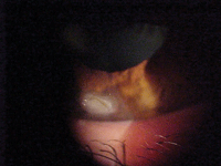
Biomicroscopy image of our 59-year-old patient who wanted a new spectacle prescription. What do you notice?
Additional testing included a more extensive patient history. He had no explanation for the lesion and could not recall any specific incident at home or work when this might have happened. Other testing might include sodium fluorescein staining; lesion probing to rule out foreign matter; Seidel testing to rule full-thickness penetration; aqueous inspection to rule out penetrating foreign body; iris inspection for “peaked” pupil (indicative of iris penetration); lens inspection, vitreous inspection and retinal inspection to rule out a foreign body insult.
The diagnosis in this case is corneal laceration of the left eye. The corneal flap appeared white because it was necrotic. So, we removed it with a curved scissor under the guidance of a corneal specialist. The lesion was probed for foreign debris and the wound was lavaged. Both eyelids were everted to rule out the presence of any foreign material or evidence of previous episodes, and a topical antibiotic was instilled in the office.
The patient was appropriately cyclopleged and placed on a topical 4th generation fluoroquinolone q.i.d. and a topical nonsteroidal anti-inflammatory preparation q.i.d. O.S.1-11 Because the patient was not in distress, topical lubricants were used q.i.d. in combination with oral analgesics for pain (as needed). We determined that a pressure patch or bandage contact lens was not necessary.8,12 We scheduled the patient for follow-up in 24 hours.4
After one week, we added a topical steroid q.i.d. to quiet corneal and scleral inflammation as well as reduce the risk of scar formation.13 The patient returned periodically during the next three weeks and exhibited an uncomplicated healing course.
While there was some corneal opacity present with a mild indentation at the site of the injury (fluorescein pooling, not staining), the epithelium filled in nicely and there was no residual effect on his vision.
At the final follow-up visit, we refracted him and updated his spectacle prescription. We also asked the patient to continue using sodium chloride ointment at bedtime O.S. for one month to reduce his risk of recurrent corneal erosion.14-16
1. Fujikawa LS, Nussenblatt RB. Recurrent and Chronic Corneal Epithelial Defects. In: Abbott RL. Surgical Intervention in Corneal and External Diseases. New York: Grune and Stratton Inc; 1987:59-67.
2. Kirkwood BJ. Recurrent corneal erosion: characteristics and management options. Insight. 2007 Oct-Dec;32(4):14-7; quiz 18-9.
3. Das S, Seitz B. Recurrent corneal erosion syndrome. Surv Ophthalmol. 2008 Jan-Feb;53(1):3-15.
4. Hall JR. Mechanical Corneal Injuries. In: Nyman JS. Problems in Optometry: Ocular Emergencies. Philadelphia: J.B. Lippincott Co.; 1990:32-44.
5. Binder, PS, Wickham GM, Zavala EY, et al. Corneal Anatomy and Wound Healing. In: Barraquer JI, Binder PS, Buxton JN, et al. Symposium on Medical and Surgical Diseases of the Cornea. St. Louis; CV Mosby Company; 1980: 1-35.
6. François J. Recurrent dystrophic erosion of the corneal epithelium. Ophthalmologica. 1978;177(3):121-33.
7. Rosenberg ME, Tervo TM, Petroll WM, et al. In vivo confocal microscopy of patients with corneal recurrent erosion syndrome or epithelial basement membrane dystrophy. Ophthalmology. 2000 Mar;107(3):565-73.
8. Kaiser PK. A comparison of pressure patching versus no patching for corneal abrasions due to trauma or foreign body removal. Corneal Abrasion Patching Study Group. Ophthalmology. 1995 Dec;102(12):1936-42.
9. Ta CN, Chan I, Dhatt HS, et al. Prospective comparison of topical moxifloxacin in eliminating conjunctival bacterial flora following a one-day or one-hour application. J Ocul Pharmacol Ther. 2008 Aug;24(4):427-31.
10. Moshirfar M, Chew J, Werner L et al. Comparison of the effects of fourth-generation fluoroquinolones on corneal re-epithelialization in rabbit eyes. Graefes Arch Clin Exp Ophthalmol. 2008; 246(10):1455-61.
11. Kaiser PK, Pineda R. A study of topical nonsteroidal anti-inflammatory drops and no pressure patching in the treatment of corneal abrasions. Corneal Abrasion Patching Study Group. Ophthalmology. 1997 Aug;104(8):1353-9.
12. Hirst LW. Pressure patching for corneal abrasions. Ophthalmology. 1997 Feb;104(2):169; author reply 170.
13. Webster RG. Corneal Injuries. In: Smolin G, Thoft RA. The Cornea. Boston: Little, Brown and Company; 1987:517-27.
14. Wang L, Tsang H, Coroneo M. Treatment of recurrent corneal erosion syndrome using the combination of oral doxycycline and topical corticosteroid. Clin Experiment Ophthalmol. 2008 Jan-Feb;36(1):8-12.
15. Smith VA, Khan-Lim D, Anderson L, et al. Does orally administered doxycycline reach the tear film? Br J Ophthalmol. 2008 Jun;92(6):856-9.
16. Arbour JD, Brunette I, Boisjoly HM, et al. Should we patch corneal erosions? Arch Ophthalmol. 1997 Mar;115(3):313-7.
