This year’s ARVO research sought to reduce the incidence of microbial keratitis, by delving deeper into microbial colonization of lens cases—like those seen here.
 Researchers offered new contact lens insights that focused on lens/storage case-induced contamination, the influence of mechanical force, and other important matters. Studies also quantified the effects of dry eye on ocular tissue, investigated meibomian gland dysfunction, and evaluated new corneal procedures.
New Data on Contact Lens Wear
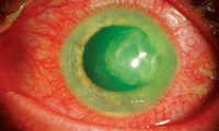
Lack of contact lens case hygiene has often been shown in epidemiology research to be associated with increased risk of microbial keratitis. The aim of yet two more trials was to examine the microbial colonization of lens cases during the use of lens care products containing dual disinfectants.693/A4
A pair of non-contemporaneous, prospective, single-group, bilaterally designed, open-labeled clinical studies employed the same protocol to evaluate the microbial contamination of contact lens cases when used with balafilcon A lenses in conjunction with RevitaLens OcuTec (polyquaterium-1 and Alexidine, Abbott Medical Optics) in one study or in conjunction with Biotrue (polyquaternium-1 and PHMB, Bausch + Lomb) in another study. Forty subjects were enrolled in each of the studies, and lens cases (approximately 70 from each trial) were collected after one month of use. The cases were cultured with the use of standard techniques to identify microbes.
Use of lens care products containing polyquaterium-1 and Alexidine or polyquaternium-1 and PHMB resulted in a low frequency of contamination of contact lens cases by gram-negative bacteria, suggesting these lens care products may help reduce the incidence of a primary source of bacterial contamination during contact lens wear.
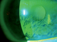
In other contact lens research, it was found that commercially available scleral lenses can help manage many conditions that compromise the ocular surface.4715/A26 Although the primary goal of scleral lens wear in the studied patient population was ocular surface protection, the researchers also found that visual acuity improved for most in the group. The research focused on the use of the Jupiter Scleral Lens (Visionary Optics/Essilor Contact Lenses).
Rigid gas-permeable scleral lenses rest entirely upon the sclera and completely vault the cornea and limbus, maintaining a fluid reservoir between the lens and the cornea. These unique fitting characteristics allow the devices to protect the ocular surface, the researchers reported. Data were collected at the Mayo Clinic in Rochester, Minn., through a retrospective chart review of patients with ocular surface disease who were fitted with these lenses between June 2006 and November 2011. The scleral lenses had been prescribed for 114 patients (185 eyes). Lenses were prescribed for assorted reasons, including chronic graft versus host disease, exposure keratopathy, neurotrophic keratopathy, limbal stem cell deficiency and Sjögren’s syndrome, to name a few.
Scleral lenses rest entirely upon the sclera and completely vault the cornea and limbus, maintaining a fluid reservoir between the lens and the cornea. These unique fitting characteristics allow the devices to protect the ocular surface.
After patients wear any type of contact lenses, notable limbal and scleral changes are known to occur. A study at the School of Optometry, Queensland University of Technology, Brisbane, Australia, used anterior segment optical coherence tomography (AS-OCT) to evaluate these changes.4729/A40 Nasal and temporal horizontal 5mm B-scans (centered on the limbus) were taken for six subjects using a commercial AS-OCT before and after six hours of contact lens wear for three types of lenses (hydrogel sphere, silicone hydrogel sphere and silicone hydrogel toric). At least one day of no contact lens wear was introduced between periods of lens wear.
Significant changes in the limbal/scleral region were observed after contact lens wear, while only limited corneal changes were observed. This limited study showed that AS-OCT can potentially be used as a powerful tool for observing the effects of contact lenses on the ocular surface.
Effects of Mechanical Force
The influences of hypoxia and mechanical force on corneal sensitivity during contact lens wear are currently unclear. In Sydney, Australia, investigators looked into the short-term influence of mechanical force exerted by contact lenses on central corneal sensitivity. Eighteen patients (seven males, 11 females, ages 23.6±4.8 years old) wore silicone hydrogel (SCL), rigid (RGP) or orthokeratology (OK) contact lenses. Lenses were matched in Dk/t (46 ISO units), but assumed to exert different levels of mechanical force on the corneal surface. The lenses were worn for a single overnight wear (eight hours) in the right eye only.
Central corneal sensitivity was found to be reduced after a single overnight wear of OK lenses, as measured using the Cochet-Bonnet aesthesiometer. This finding suggested that the mechanical force exerted by contact lenses may be a key influence on corneal sensitivity. It also indicated that Cochet-Bonnet and non-contact corneal aesthesiometer instruments may measure different aspects of corneal sensitivity.
Extended wear patients may be able to reduce risks of complications by faithfully cleaning their lenses each morning, another study found. The retrospective analysis in Sydney, Australia, and Hyderabad, India, was designed to determine if cleaning or replacing lenses in the morning or evening—during continuous wear—influenced the rate of corneal erosions.6092/D913
Continuous contact lens wear related to ocular mechanical adverse events could be reduced by changes in cleaning or replacement modality, researchers found. Changes could also be related to elimination of the overnight debris accumulation achieved through a morning replacement/cleaning modality.
In another study, researchers in Boston and Iowa City, Iowa, used laser in vivo confocal microscopy to evaluate subclinical immune responses to various contact lenses and contact lens solutions.6112/D933 The multicenter, randomized, double-blinded, clinical trial focused on 65 naive contact wearers (130 eyes) who had been fitted with silicone hydrogel contact lenses (PureVision [Bausch + Lomb], Oasys [Vistakon] or Biofinity [CooperVision]) and who had been enrolled into one of three contact lens solution groups (OPTI-FREE RepleniSH [Alcon], Clear Care [Alcon] or ReNu MultiPlus [Bausch + Lomb]).
Increased ocular surface staining and minimal ocular injection were observed in all groups. Conjunctival staining correlated to peripheral dendritic cell density (r>0.40 for all quadrants).
ReNu MultiPlus demonstrated significantly increased staining for all conjunctival, limbal and cornea areas, correlating with the highest increase in dendritic cell density in the central cornea (58%) and nasal (26%) and temporal (24%) quadrants at six weeks, compared to OPTI-FREE RepleniSH and Clear Care. Laser in vivo confocal miscroscopy revealed increased immune cell infiltration in all groups after one week of contact lens wear. Corneal and conjunctival staining were detected after six weeks of wear.
Dry Eye Advances
In an effort to create new treatments for patients with ocular surface disease, protein biomarkers for dry eye were identified in post-menopausal women.
Protein biomarkers for dry eye disease were identified in post-menopausal women by using label-free quantitative proteomics.413 Investigators from Ohio State University School of Optometry and the College of Optometry, University of Texas, tried this approach to analyze protein extracted from the Schirmer strips from normal patients (n=25) and patients with post-menopausal dry eye (n=25). Each sample was individually digested with trypsin and then analyzed by liquid chromatography–mass spectrometry.
Approximately 400 unique proteins were identified. Proteins of interest—lysozyme, lipocalin and mammoglobin B—demonstrated numerous functions and protective roles, including front-line defense, tear film stability, lipid scavengers, and products of inflammation.
In another study at the School of Optometry, Indiana University, and University of Waterloo, Ontario, Canada, researchers sought evidence that fluorescent dye quenched the tear film and affected its interpretation.4258/A135 They measured changes in tear film fluorescence over time during tear break-up (TBU) and thinning, along with associated sensory descriptors in 16 patients with a range of dry eye symptoms.
In each patient, one eye was kept open as long as possible, while the patient indicated level of discomfort on a 0 to 10 scale and described sensations. An arbitrary cutoff of 5% TBU at the end of each trial was used to divide subjects into two groups—TBU vs. tear thinning.
A decrease in tear film fluorescence over time was best explained by the phenomenon of fluorescent dye quenching as the tear film thinned because of evaporation. However, the rate of change in fluorescence was much higher when associated with rapid TBU. Associated sensations were greater, suggesting that TBU and thinning operate by different mechanisms and exert different levels of stress on the ocular surface.
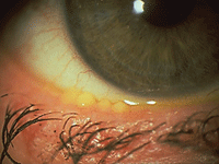
Meibomian Gland Dysfunction
One study in Boston focused on the host immune response and effects of anti-inflammatory treatment in meibomian gland dysfunction.593/A61 Laser in vivo confocal microscopy was used to analyze images of the palpebral conjunctiva and meibomian glands of one eye of five healthy individuals and both eyes of 11 patients with a clinical diagnosis of meibomian gland dysfunction. Anti-inflammatory treatment was prescribed based on laser in vivo confocal microscopy findings, resulting in follow-up visits for six patients.
Confocal microscopy indicated that meibomian gland dysfunction was associated with increased conjunctival and intraglandular inflammation. Anti-inflammatory therapy improved both clinical signs and imaging parameters, but it was followed by a lag in symptomatic relief.
Searching for causes of meibomian gland dysfunction, investigators in Tampa, Fla., identified fibrotic obstruction as a major factor.605/A73 Intraductal meibomian gland probing was designed to evaluate frequency of a popping noise and tactile relief of intraductal resistance. Gritty sensations were also associated with keratinized cell debris believed to be contributing to obstructive meibomian gland dysfunction. Of 15,642 glands probed, 41% involved popping, 24% involved gritty sensations and 35% exhibited neither of these characteristics. Up to 74% of upper glands and 50% of lower glands revealed some degree of detectable intraductal resistance.
Several studies investigated MGD, including its dignosis, cause and
treatments.
Experts at the University of Texas took a retrospective look at the effectiveness of long-term treatments of meibomian gland dysfunction.608/A76 Their study of 82 patients showed that the current treatment approach had a positive impact on patients’ signs and symptoms. Current treatments are aimed at unplugging the meibomian glands, altering meibum, and controlling inflammation on a short-term basis, typically during a period of days to weeks. Efficacy of the treatments was assessed by basal tear testing and ratings of 0 to 4 (0=none, 4=severe) for anterior blepharitis, lid margin vascularity, meibomian gland obstruction and meibum turbidity.
Statistically significant improvements were noted for anterior blepharitis and meibomian glands at all time points during the two-year study period. Anterior blepharitis scores improved by 0.79 and 0.78 points after three months and by 1.00 and 0.92 in the three-to-six-month follow-up group. Meibomian gland obstruction improved by 0.68 in the lower left lid in the zero-to-three month month group at 1.43 and at 1.86 points after the first year of follow up.
Updates on Corneal Procedures
Debate persists over whether graft thickness determines functional outcome in posterior lamellar surgery. One important study that shed new light on the subject compared descemet membrane endothelial keratoplasty (DMEK) outcomes to descemet stripping automated endothelial keratoplasty (DSAEK) at the University of Erlangen-Nuremberg, Erlangen, Germany.3137
The study looked at a single-center, retrospective, consecutive case series of 38 consecutive patients undergoing DMEK and 35 consecutive patients undergoing DSAEK for Fuchs endothelial dystrophy or pseudophakic bullous keratopathy. Three months after DMEK, 83% of the eyes reached a visual acuity of 20/40 or better, which increased to 95% six months after surgery. Thirty-six percent of eyes reached a visual acuity of 20/25 or better three months after DMEK. This proportion increased to 50% six months after surgery.
Meanwhile, three months after DSAEK, 28% of eyes reached a visual acuity of 20/40 or better, which increased to 43% six months after surgery. Therefore, DMEK was found to provide faster and more complete visual rehabilitation six months after surgery when compared to DSAEK. Endothelial cell survival was not significantly different between either group, within a six-month follow-up period.
Researchers in Portland, Ore., used anterior corneal topography to measure improvements in anterior corneal topography for up to two years after DSAEK.3139 From a prospective data registry of corneal transplant recipients, researchers identified 75 eyes of 58 patients who received DSAEK for Fuchs’ corneal endothelial dystrophy or pseudophakic bullous keratopathy. These patients had no ocular comorbidities known to limit visual acuity and had complete datasets for three years of post-operative follow-up.
Best spectacle-corrected visual acuity improved at each time point, but not every interval revealed statistical significance. Mean Snellen best corrected visual acuity was 20/51 pre-operatively, 20/30 at six months, 20/29 at 12 months, 20/25 at 24 months, and 20/24 at 36 months.
Researchers studied ReLEx smile (Carl Zeiss Meditec AG), a new femtosecond laser-based, small-incision lenticule extraction procedure that addresses moderate and high myopia.5575 A total of 379 eyes (198 patients) were treated for myopia (spherical equivalent ranging from -13.13D to -1.63D, mean -7.28D) with ReLEx smile and prospectively followed for three months.
ReLEx smile is a flapless, all-in-one refractive procedure. Refractive predictability, safety and patient satisfaction at three months seemed equal to ReLEx flex and femtosecond-LASIK. Optimizing laser energy settings and surgeon experience was confirmed to be important to minimize initial inferior results.
