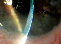
 Q: I recently saw a patient with a peculiar bilateral peripheral corneal edema and a clinical history that included a past intracapsular surgery for cataracts (now pseudophakic). I sent the patient for a corneal consult and the diagnosis was Brown-McLean syndrome. What is this entity and what causes it? Are there any effective treatments?
A: First described in 1969, Brown-McLean syndrome (BMS) is a clinical entity of peripheral circumferential corneal edema that usually starts in the inferior cornea and is often associated with orange-brown pigmentation at the level of Descemet’s membrane.1 Because the central cornea is usually spared, vision is often normal.
This condition is frequently asymptomatic, but the most common presenting symptom is foreign body sensation from bulla formation and, in some instances, pain due to ruptured bullae.1 The cause of BMS is still unknown, but researchers suggest it may develop in patients who have a genetic predisposition when their eyes are exposed to certain conditions, such as the insertion of an anterior chamber lens.2
“It is most commonly seen after intracapsular cataract extraction, although it can be seen after any intraocular procedure, and occasionally in patients who have never had surgery,” says Thomas S. Boland, M.D., of Northeastern Eye Institute in Scranton, Pa.

Brown-McLean peripheral corneal edema in an eye with long-standing aphakia. Photo: Alan Sugar, M.D.
BMS has been linked to several lens surgeries, including extracapsular lens extraction, phacoemulsification, pars plana lensectomy and vitrectomy.1
The onset of this condition usually ranges from six to 16 years after the time of procedure.2 “Due to the decline of intracapsular surgery over the last 20 to 30 years, it is rarely seen these days,” Dr. Boland says. “It can usually be treated conservatively with lubrication, hyperosmotics and monitoring for infection in the case of ruptured bullae.” In some cases, anterior stromal puncture or a bandage contact lens have been suggested as alternative treatments to control severe foreign-body sensation.3
It’s important to educate patients with BMS about the signs and symptoms of corneal surface problems, particularly corneal ulceration secondary to contact lens wear, and to perform regular follow-up to monitor for any disease progression. Some clinicians have found confocal microscopy to be a convenient and informative means of documenting BMS as well as a useful tool in follow-up.4
1. Gothard TW, Hardten DR, Lane SS, et al. Clinical findings in Brown-McLean syndrome. Am J Ophthalmol. 1993 June 15;115(6):729-37.
2. Pareja-Esteban J, Montes MA, Perez-Rico C, et al. Brown-McLean syndrome after insertion of an anterior chamber intraocular lens: description of one case. Arch Soc Esp Oftalmol. 2007 May;82(5):315-7.
3. Martins EN, Alvarenga LS, Sousa LB, et al. Anterior stromal puncture in Brown-McLean syndrome. J Cataract Refract Surg. 2004 Jul;30(7):1575-7.
4. Lim LT, Tarafdar S, Collins CE, et al. Corneal endothelium in Brown-McLean syndrome: in-vivo confocal microscopy finding. Semin Ophthalmol. 2012 Jan-Mar;27(1-2):6-7.

