 In late December 2011, a 70-year-old white male presented to the office as a new patient to establish care. He had moved to the area approximately six months earlier and needed to have his glaucoma medications refilled. These included Lumigan (bimatoprost, Allergan) h.s. O.U., Azopt (brinzolamide, Alcon) b.i.d. O.U. and Alphagan P (brimonidine, Allergan) 0.1% b.i.d. O.U. He began glaucoma therapy approximately eight years earlier.
His systemic medications included lisinopril, Zetia (ezetimibe, Merck/Schering-Plough), atenolol, Plavix (clopidogrel, Bristol-Myers Squibb/Sanofi), low-dose aspirin (81mg), Cardizem (diltiazem, Valeant Pharmaceuticals/Abbott Laboratories), Nexium (esomeprazole, AstraZeneca), amiodarone and Arthrotec (diclofenac/misoprostol, Pfizer) b.i.d. He reported an allergy to penicillins.
Significant in his medical history was coronary artery bypass graft (CABG) surgery (x3) approximately 10 years earlier, and a left carotid endarterectomy shortly thereafter.
Diagnostic Data
At his initial visit, best-corrected visual acuity was 20/25- O.D. and 20/30- O.S. through myopic astigmatic correction. Pupils were equal, round and reactive to light and accommodation, with no afferent defect. Extraocular motilities were full in all positions of gaze. Confrontation fields were full O.U.
Slit lamp examination of the anterior segment was only remarkable for corneal arcus in both eyes. The anterior chamber was deep and quiet, and angles measured by Van Herick estimation were grade 3- open in both eyes. Intraocular pressure measured 8mm Hg O.D. and 10mm Hg O.S. at 9:45 a.m. Pachymetry readings were 571μm O.D. and 563μm O.S.
Dilated fundus exam showed crystalline lenses with moderate nuclear sclerosis in both eyes, with cortical cataracts encroaching on the visual axis (O.S. > O.D.), consistent with his best-corrected visual acuity. Posterior vitreous detachments were present bilaterally.
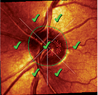
Stereoscopic evaluation of his optic nerves demonstrated cup-to-disc ratios of 0.65 x 0.65 O.D. and 0.50 x 0.50 O.S. The neuroretinal rims did not respect the ISNT (inferior-superior-nasal-temporal) rule in either eye. Retinal vasculature was characterized by moderate arteriolar sclerosis in both eyes with scattered arteriovenous crossing changes. The retinal venules were mildly dilated, but no overt evidence of occlusive disease was present––including retinal hemorrhages, micro-infarcts or areas of retinal ischemia or hypoxia. Both maculae were characterized by fine retinal pigment epithelial granulation consistent with his age. The peripheral retinal evaluations in both eyes were unremarkable. I obtained stereo-optic nerve photos at this visit.
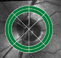
Heidelberg Structure and Function printout of the patient’s left eye shows a normal optic nerve Moorfields Regression Analysis (middle left and inner green circle, left image) and a normal HEP field (middle right and outer green circle, right image). So, why was he on three glaucoma drops?
We did not have the time to do a complete glaucoma evaluation at this initial visit, so I refilled his three glaucoma medications and asked him to return in a month for further glaucoma testing. In the interim, I sought to obtain his previous records to ascertain the chain of events that led to him being medicated for IOP of 10mm Hg and below.
In January 2012, the patient returned as directed. At this visit, we administered Heidelberg Edge Perimeter (HEP, Heidelberg Engineering) visual field testing in both eyes, which were normal with good reliability. IOP at this visit measured 9mm Hg O.D. and O.S. at 11:00 a.m.
Gonioscopy revealed open angles in both eyes with minimal trabecular pigmentation and no ciliary body visible. Angle anatomy was normal with no plateau iris configuration, peripheral anterior synechiae or other abnormalities. Ultrasonic B-scan of the anterior chamber angles confirmed normal iridocorneal angle anatomy and normal posterior chambers with normal iris configuration.
Heidelberg Retina Tomograph-3 (HRT-3, Heidelberg Engineering) imaging of both optic nerves confirmed my earlier description: relatively plush, average-sized neuroretinal rims that did not adhere to the ISNT rule. Moorfields Regression Analysis did not identify any sector of either optic nerve with statistically aberrant parameters.
Discussion
By this point, I had seen the patient twice; in both instances, I did not pick up on one piece of evidence that he had frank glaucoma (except that he has been medicated with three different drugs). His optic nerves looked healthy with moderate cupping. Selective perimetry was normal in both eyes, and medicated IOPs were in the single digits. I could come up with only a few rationales for his therapy with three glaucoma drops:
- Perhaps when he was first placed on medications years ago, he was an ocular hypertensive patient with IOPs elevated enough to prompt the treating physician to drive the pressures down very low.
- Or, his history of cardiovascular disease, retinal arteriolar sclerosis and carotid artery disease may have given the previous provider the sense that ocular perfusion pressure to the eye(s) was low, which prompted prophylactic IOP lowering.
In reality, it may have been a combination of both factors that ultimately led him to be prescribed three glaucoma medications.
Whether he would be best served with a laser procedure to reduce the number of medications he is currently taking is not of concern here. The primary concern lies with the unanswered question as to why he is even on medication.
Fortunately, his past records arrived prior to his second visit. In reviewing these records, it appeared that initial IOP readings were in the low to mid-20s, as measured by non-contact tonometry on some occasions and by Tono-Pen (Reichert) on other occasions. IOP never exceeded 24mm Hg in either eye.
Furthermore, his records noted “multiple CV issues” on a few of the visits. Reading between the lines, my guess is that the previous provider may have been concerned with blood flow issues to the eyes; his initial visits to the previous eye care provider occurred shortly after his CABG and endarterectomy surgeries.
Now What?
The question now remains: How should we proceed? While there may be some benefit to ameliorating the risks of retinal vascular occlusion (in this case, reducing the risk of retinal vein occlusions) by lowering IOP, I do not think he needs to have his IOP lowered to prevent glaucomatous damage, as he apparently is an ocular hypertensive patient.
After discussing my findings and thoughts with the patient, I relayed to him my strong feelings about keeping patients on the fewest number of medications that “get the job done.” Accordingly, I asked him to discontinue the Alphagan. He did so, and on follow-up in late March 2012, IOP measured 14mm Hg O.D. and 13mm Hg O.S. Optic nerves remained stable, as did the retinal vasculature. Continuing on the same thought process, I asked him at that visit to discontinue Azopt, and see me again in four to six weeks.
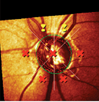
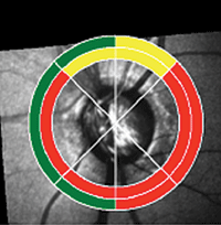
A different patient showing structural and coincident functional deficits on one
printout, as measured by the HRT-3 and HEP.
When last seen in early May 2012, his IOP measured 18mm Hg O.D. and O.S. at 9:45 a.m., with stable optic nerves, fields and retinal vasculature. While IOP is at an acceptable range for an ocular hypertensive with slightly thick corneas and normal optic nerves, it remains to be seen whether all will remain stable over time.
Underdiagnosis and Overdiagnosis
Given that we live in a very mobile society and that Americans move to different locations of the country more now than generations ago, we will be seeing more and more patients who need continuing care. And, when we see new patients, we will sometimes see individuals who are undiagnosed or overdiagnosed.
Interestingly, a recent study looked at Medicare claims in various regions of the country among patients who carried a diagnosis of glaucoma or glaucoma suspect.1 The study found that, in certain areas of the country (the Northeast corridor and Mid-Atlantic states), there is a higher likelihood of being diagnosed with either of those, whereas in other parts of the country (such as the South and Southeast), the likelihood of being diagnosed with glaucoma or as a glaucoma suspect is much lower.1
It begs the question: In those areas where diagnosis rates are higher, are they higher because the providers in those areas do a better job of making the diagnoses, or is it because providers in those areas tend to overdiagnose their patients? And, what role does the variability in frequency of patient visits play in these differing diagnoses rates? These questions require further investigation.
In the interim, each of us is charged with rendering an opinion of our patient’s status. Of course, we should take an objective look at what the previous provider (if there was one) was seeing and thinking when managing the patient. But, our primary goal is to offer the patient the care that we think is best for them––whether that corresponds with earlier treatment, or contradicts it.
1. Cassard SD, Quigley HA, Gower EW, et al. Regional variations and trends in the prevalence of diagnosed glaucoma in the Medicare population. Ophthalmology. 2012 Apr 3. [Epub ahead of print]
