 In 2009, a 41-year-old white female presented as a new patient with complaints of transient blurred vision (O.S. > O.D.) and transient headaches that had persisted for the previous two months. She reported that the visual blur tended to wax and wane throughout the day, but would generally affect her vision for a period of four to five days, then clear for as long as two to three days at a time.
During the episodes of visual blur, the patient also noted some periocular headaches that tended to be worse in the mornings; these too waxed and waned, and did not always coincide with the blurred vision.
Two months prior to this visit, she had seen her primary care provider who had diagnosed her with sinusitis and sinus-related headaches following a CT scan. After several trials of medications to alleviate both, she eventually found that Claritin (loratadine, Schering-Plough) seemed to give her the most relief with her headaches and also seemed to reduce the frequency of visual blur.
Then her headaches and blurred vision began to worsen in frequency and intensity. She also reported feeling “lightheaded.” Given the progressive symptoms, her primary care physician referred her to me for an evaluation.
Diagnostic Data
Her medications included Claritin and Ortho Tri-Cyclen (norgestimate/ethinyl estradiol, Ortho-McNeil-Janssen Pharmaceu-ticals). She reported no known allergies to medications.
Best-corrected visual acuity was 20/20 O.D. and O.S. Pupils were equal, round and reactive to light and accommodation with no afferent defect O.U. Extraocular motilities were full in all positions of gaze and Amsler grid testing was negative O.U. Color vision was normal. Threshold visual fields demonstrated a completely normal field O.D., and a slightly enlarged blind spot O.S.
Slit lamp examination of her anterior segments was unremarkable. Anterior chamber angles appeared grade IV open. Intraocular pressure measured 14mm Hg O.D. and 15mm Hg O.S.
Upon dilated examination, her crystalline lenses were clear. Stereoscopic examination of her optic discs revealed bilateral elevated disc margins, as well as a disc hemorrhage O.D. at one o’clock. Otherwise, her retinas appeared normal.
Given her apparently clear CT scan (except for the sinusitis) just one month earlier, a space-occupying lesion was not the likely cause of her symptoms. I considered several differentials, including venous sinus thrombosis and pseudotumor cerebri (PTC), given that she was mildly obese.
Consequently, we ordered an MRI, MRA and MRV, which were all found to be normal. The MRI demonstrated an empty sella. Lumbar puncture demonstrated elevated opening pressures.
Diagnosis and Management
Ultimately, I diagnosed the patient with PTC—now termed idiopathic intracranial hypertension—because she met the Modified Dandy Criteria.1,2 (Recall that these criteria include signs and symptoms of increased intracranial pressure as well as elevated cerebrospinal fluid pressure, among others.)
IOP isn’t the only pressure to consider. Cerebrospinal fluid pressure also exerts force on the optic nerve and the posterior lamina cribrosa. Image: Intracranial Hypertension Research Foundation,
www.ihrfoundation.org
Subsequently, she successfully managed the condition with weight loss, exercise and acetazolamide on a titrated scale.
But recently, during a regular follow-up visit in September 2012, she reported that her mother and both of her siblings had been diagnosed with glaucoma, and she was concerned that glaucoma would pose a threat to her vision, too.
So, how do you counsel such a patient? Given there is no clear evidence that she has glaucoma—nor even that she is a suspect—glaucoma is not a relevant issue for her at this time.
But, is there a link between the development of glaucoma and intracranial pressure?
Discussion
In the past few years, research has looked more closely at the relationship between intracranial pressure, as measured by cerebrospinal fluid pressure (CSFp), and the development of glaucoma.
One of the newer terms that has been articulated and used more regularly is the so-called translaminar pressure gradient (TLPG). As its name implies, TLPG is the pressure differential across the lamina cribrosa between the intraocular pressure and the CSFp in the optic nerve subarachnoid space.
Interestingly, several cases of bilateral disc edema in astronauts returning from prolonged visits to the International Space Station has brought greater attention to TLPG.3 Also, some researchers have opined that the role of CSFp and its relationship to IOP may have a more direct role in the development of glaucoma than previously thought.4
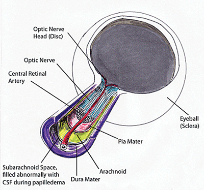
From an anatomical perspective, remember that the optic nerve travels through two separate pressurized anatomical regions. First, we all know about the IOP’s effect on the intraocular portion of the optic nerve. But we also need to keep in mind that the retrolaminar portion of the optic nerve (that part of the optic nerve posterior to the lamina cribrosa in the orbit and suprasellar cistern) is enclosed in meninges through which CSF travels. So, not only is part of the optic nerve affected by IOP in the globe, but a larger and longer portion of the optic nerve is surrounded by CSF—a portion called the optic nerve subarachnoid space (ONSAS).
Keep in mind that the lamina cribrosa is essentially sandwiched between the IOP (anteriorly) and the ONSAS (posteriorly). So, it makes sense that a substantial pressure differential between the IOP anteriorly and the CSFp posteriorly can have a significant effect on the optic nerve in the laminar region. A relatively low CSFp at the level of the ONSAS may likely predispose a person to develop glaucoma, because the relatively higher IOP can push against the lamina with little posterior “support.” If this is true, then it also stands to reason that a slightly elevated CSFp in the ONSAS may help protect against glaucomatous optic nerve damage.
But several questions remain. If the most readily and traditionally used measure of CSFp is through a lumbar puncture, then does lumbar puncture entering pressure equate to CSFp in the optic nerve? That’s a fair question, and while measuring CSFp in the ONSAS is difficult, visualization of the ONSAS is relatively easy with high-resolution MR techniques.
So, researchers recently looked at the width of the optic nerve subarachnoid space (ONSASW) as a substitute for measuring CSFp in the ONSAS—particularly in terms of the relationship between glaucoma and the orbital optic nerve CSFp.5 Specifically, they looked at the ONSASW in groups of POAG patients with normal and elevated IOP, as well as in a control group. Interestingly, they found that in the normal-pressure POAG group, the ONSASW was significantly narrower than in patients with high-pressure POAG as well as those in the control group. They concluded that a narrower ONSASW is consistent with a lower CSFp in the ONSAS, and that this lower CSFp in the ONSAS may correlate with a higher likelihood of normal-pressure glaucoma.
Many questions still need to be answered, but it does appear that CSFp—or at least the orbital portion of the CSFp—may very well play a role in the likelihood of developing glaucoma, and perhaps more so in those with normal pressure glaucoma.
So, what about our patient with PTC? She had asked whether she should be concerned about developing glaucoma. Will a simple answer—“No, there’s no problem with glaucoma in your case”—be enough for her? Probably so.
But is that enough of an answer for us as providers of glaucoma care? No, it’s not, because we have yet to understand the full relationship between CSFp and IOP.
1. Smith JL. Whence pseudotumor cerebri? J Clin Neuroophthalmol. 1985 Mar;5(1):55-6.
2. Friedman DI, Jacobson DM. Diagnostic criteria for idiopathic intracranial hypertension. Neurology. 2002 Nov 26;59(10):1492-5.
3. Mader TH, Gibson CR, Pass AF, et al. Optic disc edema, globe flattening, choroidal folds, and hyperopic shifts observed in astronauts after long-duration space flight. Ophthalmology. 2011 Oct;118(10):2058-69.
4. Berdahl JP, Allingham RR. Intracranial pressure and glaucoma. Curr Opin Ophthalmol. 2010 Mar;21(2):106-11.
5. Wang N, Xie X, Yang D, et al. Orbital cerebrospinal fluid space in glaucoma: The Beijing Intracranial and Intraocular Pressure (iCOP) Study. Ophthalmology. 2012 Oct;119(10):2065-2073.e1.
