 |
A 60-year-old African American male presented complaining of a red, irritated right eye with associated brow ache, and he felt his vision had decreased slightly in that eye.
He reported a history of glaucoma surgery (trabeculectomy) in both eyes approximately five years earlier and was not on any topical glaucoma medication currently. His uncorrected distance visual acuity was 20/50 OD and 20/25 OS, and pinhole acuity was 20/30 OD.
Slit-lamp examination revealed a superiorly located whitish bleb surrounded by 3+ conjunctival injection. There appeared to be a mucopurulent infiltrate within the bleb in the right eye. The left eye showed a patent bleb that was relatively avascular, and the conjunctiva was quiet. The anterior chamber was deep, showing trace cell and flare, in the right eye.
Intraocular pressure (IOP) was 5mm Hg OD and 9mm Hg OS. NaFL strip application revealed a subtle leak from the infero-lateral edge of the bleb in the right eye. Dilated examination revealed advanced cupping in both eyes.
The periphery was intact without choroidal effusions. Vitreous cells were not evident on slit-lamp exam or under binocular indirect ophthalmoscopy. B-scan ultrasound was acoustically normal in both eyes.
Discussion
One of the most concerning complications after glaucoma filtering surgery is infection.
HELP, an acronym for “hypotony, endophthalmitis, leak and pain,” is used to assist clinicians in remembering the key signs and symptoms associated with blebitis or bleb-related endophthalmitis, urgent complications of trabeculectomy.
It is also important to differentiate between blebitis and bleb-related endophthalmitis, as each has distinct management, treatment and prognosis.
Patient Education
Urge surgical patients to remember their own acronym, RSVP, “redness, sensitivity, vision loss and pain.” If these symptoms develop, they should contact their eye doctor or surgeon immediately, no matter how long ago they had surgery.
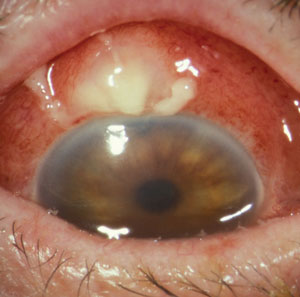 | |
| Blebitis with decreased vision and pain prior to antibiotic therapy. |
In the absence of such education, patients may not think their symptoms are related to a procedure they had years ago. Patients are more apt to link a red, irritated eye to surgery if the symptoms develop within hours, days or months of the surgery. However, in the case of bleb-related infections, average time of onset is approximately three to four years after trabeculectomy.1,2
With timely intervention, blebitis will have a much better prognosis than an infection that migrates posteriorly and evolves into bleb-related endophthalmitis.
It is also important that trabeculectomy patients and comanaging physicians be educated on the avoidable (e.g., eye rubbing) and unavoidable risk factors associated with both early- and late-onset bleb leak and infection. For example, any excessive exposure to an antimetabolite or antifibrotic agent during surgery should be communicated to the comanaging eye care practitioner. The intraoperative use of antifibrotic agents like 5-fluorouracil and mitomycin-C decrease unwanted scarring after trabulectomy surgery, thereby decreasing the risk of postsurgical bleb failure. However, these agents may weaken and thin the overlying conjunctiva, increasing the risk of a bleb-related leak or infection.3
Although there does not have to be a bleb leak for infection to occur, one case-controlled study reported that the risk of infection is 25 times greater in the presence of a late-onset bleb leak.4
Stay Alert for HELP
Eye care providers should check blebs for leakage at every visit. Some patients may be asymptomatic. Complaints of excessive tearing or watering eyes in association with even a slight IOP decrease should raise suspicion of a leak.
When assessing a bleb for potential leaks, pay attention to the thinnest, most avascular areas. It is more common to find leaks at the apex of a cyst, as these areas are most likely to dry out due to poor wetting from abnormal tear flow. Additionally, these areas are more prone to dehiscence secondary to mechanical lid rubbing.
Checking for leaks is most commonly done by applying a modest amount of sterile saline to the end of a NaFl sterile strip, followed by “painting” the thinnest apical areas of the bleb while viewing under a cobalt-blue filter at the slit-lamp biomicroscope. If there is no “Seidel sign” or evident flow of the dye, then check the base of the bleb.
Treatment and Management
Blebitis and bleb-related endophthalmitis warrant different strategies. When bleb leak is evident without signs of infection or anterior chamber involvement, initiate conservative therapy.5
In some cases, the combination of a topical fourth-generation FQ and an aqueous suppressant (i.e., topical beta-blocker) are enough to quell a small leak. If the IOP is too low to justify the aqueous suppressant, a large, soft bandage contact lens or a cyanoacrylate glue can be effective. According to one study, the use of a large diameter (17.5mm) soft bandage contact lens was effective at quelling bleb leaks, regardless of whether an antifibrotic agent was used during surgery.6 Another study reported an 80% success rate using a cyanoacrylate tissue adhesive, eliminating the need for bleb revision.7
When using a bandage contact lens, it can take anywhere from one to three weeks for a leak to seal. If these conservative measures do not quell the leak within this time frame, refer for bleb revision.
Blebitis
When confronted with a blebitis, defined as mucopurulent infiltrate identified within a bleb, aggressive treatment is warranted. If there is no evident anterior chamber response, you can use a commercial fourth generation FQ Q1H around the clock. Oral antibiotics with good ocular tissue penetration (e.g., moxifloxacin) are also justified. Monitor these patients daily, and in some instances, such as a monocular patient, twice per day. If there is an anterior chamber response (1+ cell or greater), topical fortified antibiotics are recommended. The most commonly used fortified ABs include Vancocin (vancomycin, ViroPharma) 33mg/ml plus Tobrex (tobramycin, Alcon) or gentamicin at 15mg/ml.
Bleb-related Endophthalmitis
The diagnosis changes to bleb-related endophthalmitis once one of the following is present: a hypopyon; cells in the anterior vitreous; or culture-positive aqueous or vitreous humor biopsy. If slit-lamp exam reveals a hypopyon or cells in the anterior vitreous, refer the patient to a retina specialist. B-scan ultrasonography will help confirm anterior vitreous cells and assist the specialist in monitoring responsiveness to treatment, which is likely to include a vitreous tap and injection of fortified ABs or vitrectomy.
Prognosis
Overall, the prognosis for blebitis is good. One should expect a favorable response both clinically and symptomatically within 48 hours.8 However, the diagnosis of bleb-related endophthalmitis usually means a four or more line drop in best-corrected visual acuity, with a majority of cases stabilizing at counting fingers or light perception vision.9 This is why the diagnosis of blebitis carries with it a significant responsibility for treatment adherence by the patient and close monitoring by the doctor. If you have concerns about a patient strictly adhering to Q1H dosing, hospital admission may be warranted.
Our patient was given a diagnosis of blebitis and was started on topical fortified antibiotics (Vancomycin and gentamicin) every hour around the clock for the first 24 hours, then every hour while awake and Q3H during the night for the next 48 hours. The patient was monitored daily. Thankfully, the eye responded to treatment and his vision was preserved. The bleb leak resolved spontaneously.
Filtering surgery has been a tremendous addition to the arsenal in the fight against glaucoma. But it comes with a lifetime commitment for both patient and doctor in monitoring for serious secondary complications like infection.
Is HELP on the way? Let’s hope not!
1. Allingham RR, Damji KF, Freedman S, et al. Shields’ Textbook of Glaucoma. Philadelphia: Lippincott, Williams & Wilkins; 2005.2. Kim E, Law SK, Coleman AL, et al. Trabeculectomy associated with 2% incidence of bleb infection at 10 years. Am J Ophthalmol. 2015.Jun;159:1082-91.
3. Bindlish R, Condon GP, Schlosser JD, et al. Efficacy and safety of mitomycin-c in primary trabeculectomy. Ophthalmology. 202;109:1336-42.
4. Soltau JB, Rothman RF, Budenz DL, et al. Risk factors for glaucoma filtering bleb infections. Arch Ophthalmol. 2000;118:338-42.
6. Shoham A, Tessler Z, Finkelman Y, Lifshitz T. Large soft contact lenses in the management of leaking blebs. CLAO J. 2000 Jan;26(1):37-9.
7. Zalta AH, Wieder RH. Closure of leaking filtering blebs with cyanoacrylate tissue adhesive. B J Ophthalmol. 1991;75:170-3.
8. Chen PP, Gedde SJ, Budenz DL, et al. Outpatient treatment of bleb infection. Arch Ophthalmol. 1997;115:1124-8.
9. Ciulla TA, Beck AD, Topping TM, et al. Blebitis, early endophthalmitis, and late endophthalmitis after glaucoma-filtering surgery. Ophthalmology. 1997;104:986-95.
