
Prism can be very useful when treating patients with symptomatic binocular vision disorders, but opinions vary widely about how to best determine the amount to prescribe. Indeed, common methods for determining prismatic prescriptions can result in different magnitudes of recommended prism for the same patient. This lack of consensus, along with rough guidelines and apparently contradictory rules of thumb, may make some clinicians uncomfortable prescribing prismand may even deter them for doing so.
This article, the first installment of our new series Back to the Basics, discusses the use of prism for treating cases of diplopia, symptomatic heterophorias and abnormal head postures.
Prism placement either eye Base-out (BO) over either eye Base-down (BD) over right eye Base-up (BU) over left eye BU over right eye BD over left eye
Prisms for Esotropic Diplopia
Basic Prism Placement
Comitant
deviation
Exo
Eso
Right hyper
Left hyper
Base-in (BI) over
A 46-year-old white male, a professor, presented complaining of distance diplopia that increased in frequency over time through his myopic correction of -5.75 -2.00 x 180 O.D. and -6.00 -2.00 x 180 O.S. Examination revealed intermittent alternating esotropia of 25 prism diopters (∆) to 30∆ at distance and 35∆ to 40∆ at near. Sensory testing revealed uncrossed diplopia beyond 6 feet and normal correspondence.
The patient refused to consider strabismus surgery, so we discussed such spectacle options as multiple spectacle corrections; single-vision, bifocal or progressive lenses; Fresnel prisms and ground-in prisms. The patient asked for one single-vision spectacle prescription that best balanced distance acuity with the largest range of single vision possible.
Refraction yielded -4.25 -1.75 x 002 for 20/20 O.D. and -4.75 -1.50 x 170 for 20/20 O.S.; the distance deviation was reduced to 18∆. The minimum amount of relieving prism that allowed for stable sensory fusion of most objects in the room at intermediate and some far viewing distances was 10∆ base-out (BO), split equally between the eyes.
The patient declined Fresnel prisms due to unacceptably reduced acuity and distortion, even when we placed total prism in front of the non-dominant eye. We prescribed spectacles with 10∆ BO ground-in prism.
Although the patient was satisfied with his new glasses, he opted for vision therapy in an attempt to eliminate the need for prism. Along with weekly therapy, prism was decreased by 1.0∆ to 2.0∆ steps about every two months as fusional vergence ranges increased. This patient eventually was able to wear his full distance spectacle correction without prism. Five months after his last office visit, the patient thanked us for permanently decreasing his class size by one-half, since he no longer experienced diplopia while lecturing.
Diplopia can be a debilitating consequence of decompensating binocular skills in patients who once may have had well-controlled motor fusion skills. Clinicians have recommended numerous treatmentsincluding patching, strabismus surgery, monovision, prism, Botox (botulinum toxin type A, Allergan) injections and vision therapyto alleviate the intolerable diplopia experienced by these patients.1-4
Unless you unnecessarily take away significant astigmatic correction in each eye, creating monocular diplopia, or put prism in the wrong direction, your prescription is unlikely to make the patient more symptomatic. Trial framing your prescription in free space before prescribing will prevent even this from occurring.
When patients report diplopia, particularly if recent in onset, you must first rule out potentially life-threatening etiologies. In adults, especially those with hypertension or diabetes, vasculopathic ischemic infarction is a common cause of fourth-nerve paresis. Midbrain or cerebellum tumors, aneurysm and ischemia can also cause paresis, resulting in diplopia.
Once the potentially insidious conditions are ruled out, ask the patient whether the double images are displaced horizontally, vertically or diagonally. This tells you whether you may need horizontal and vertical prism or just one type of prism to treat the patient. If the images are displaced diagonally, you may want to determine if you are dealing with a primary or secondary vertical deviation before prescribing prism, as vertical prism is not warranted for secondary vertical deviations.
Primary verticals are the same in direction and size, whether the eyes are in strabismic or non-strabismic alignment.5 Secondary verticals are only present when the eyes are in strabismic position and absent when the eyes are aligned.5 Therefore, a common misconception is to use horizontal neutralizing prism to align the eyes before looking for a vertical deviation. This is incorrect because behind the prism, the eyes are still physically misaligned, so even a secondary deviation will still manifest. So, unless the patient has an intermittent deviation in which he or she can physically align the eyes long enough for testing, determining whether a vertical deviation is primary or secondary can be quite challenging even for an experienced clinician.
Therefore, without having to get into techniques requiring specialized equipment such as disparometers, a general guideline to follow is that secondary verticals tend to be very small and primary verticals are larger. If an intermittent exotrope has a 25∆ horizontal displacement and only 2∆ or 3∆ vertically when the eyes are strabismic, this is most likely a secondary vertical, and correction with vertical prism is not necessary.
But, if the patient instead reports 8∆ to 10∆ vertical separation in addition to the horizontal deviation, he or she is unlikely to be able to align the eyes vertically even if the patient could pull his or her eyes straight horizontally. The latter is more likely to be a primary vertical, and horizontal and vertical prism will likely be required to relieve the patients symptoms.
The next step is to determine whether the diplopia is worse at distance or near, upgaze or downgaze, or in any particular field of gaze. A deviation that is the same in all fields of gaze with either eye fixating is called a comitant deviation. A deviation that varies in size from gaze to gaze or varies when the right eye fixates vs. when the left eye fixates is considered incomitant, or noncomitant. A noncomitant deviation often indicates a muscle underaction or paresis.
Rather than looking for an exact prism value when labeling a deviation comitant or noncomitant, look for a pattern in which the deviation appears to be larger when the eyes are moved in one direction and smaller when the eyes are moved in the opposite direction. When this type of pattern is seen, a paresis of one of the muscles may be responsible for pulling the eyes in that direction. Perform duction testing by covering an eye and repeat extraocular muscle testing monocularly, paying careful attention as each eye moves in the direction in which the deviation was largest. Prism neutralization in all nine gazes can also be performed. Generally, a deviation is considered comitant if the difference is less than 5∆ between the gazes.
Keep in mind that a longstanding paresis can undergo a spread of comitance. So, a once obviously noncomitant deviation becomes increasingly comitant with time, and the measured angle when the affected eye fixates becomes the same as when the sound eye fixates.6
Performing the Maddox Rod Test for Correspondence First, place the Maddox rod so that the striations are horizontal for horizontal deviations and vertical for vertical deviations. While viewing a transilluminator or penlight, patients see a single white spot of light and one vertical red line if the striations are horizontal (or a horizontal line if the striations are vertical). The line represents the eye with the Maddox rod, and the white light represents the other eye.
Once you determine comitancy, you will want to know if the patient has the potential for normal correspondence. There are several ways to perform this test without requiring expensive equipment such as an major amblyoscope. One method requires merely a Maddox rod, which may already be available as an accessory lens in your trial lens set or commonly on the other end of a cover paddle. This technique works best if you do not perform this test using the Maddox rod in the phoropter. (See Performing the Maddox Rod Test for Correspondence.)
Relative location of the white light to the line allows you to determine if the patient has crossed or uncrossed diplopia. Crossed diplopia occurs when the patient sees the right eyes image to his or her left and the left eyes image to his or her the right. Uncrossed diplopia occurs when the patient sees the corresponding image on the same side as the eye.
You must determine if the magnitude and direction of the prism that makes your patient perceive that the line bisecting the white light (subjective angle) matches the magnitude and direction of prism found with alternate cover test (objective angle). In other words, the subjective response of the patient should match your objective findings. If they do not, your patient has anomalous correspondence.
The same test described above could be administered using a red lens with vertical dissociating prism instead of a Maddox rod. The basic question that needs to be answered when determining correspondence is whether the patient thinks the eyeturn is the same as the actual eyeturn. A patient who perceives a deviation that is the same magnitude and direction as your measured value has normal correspondence. A patient who reports a deviation that is larger or smaller or in a different direction than your measured value has anomalous correspondence.
Whether testing is performed with a red lens and vertical prism or a Maddox rod, perform the test with optimal correction and at a distance to minimize accommodative influences that can affect the magnitude of the deviation. Keep testing conditions as similar as possible when determining the objective and subjective findings. In other words, do not perform subjective Maddox rod behind the phoropter and compare it to the findings from an objective cover test performed in free space. Finally, make sure the same eye is fixating during objective and subjective testing so that the difference found when a deviation is noncomitant is not mistaken for anomalous correspondence.
Treatment challenges caused by sensory anomalies, such as anomalous correspondence, suppression and amblyopia, are greater with early onset, long-standing deviations.5,6 These cases require more complicated diagnostic and treatment plans than the extent of this article.
If your patient has normal correspondence and is diplopic, one simple way to determine an acceptable amount of prism that eliminates the diplopia is to trial frame prism in free space using a prism bar or loose prism. Simply have the patient view a relatively large target, such as the 20/200 or 20/400 letter or a large object, in the distance. Add prism, starting with a low amount, and work your way up until the patient reports relatively stable, comfortable fusion of the target.
The clinical finding that a patient may need higher amounts of prism as smaller targets are used may be explained by Panums area. Panums area allows for some imprecision in ocular alignment without the perception of diplopia. The size of Panums fusional area is smallest at the fovea and increases in size as you move farther into the retinal periphery. Small targets restricted to the central area of retina will be perceived as diplopic easier than larger objects that can take advantage of the larger extent of Panums in the retinal periphery.7
Trial frame the prism and allow the patient to look around the room to determine if he or she sees single and feels comfortable, much like you would when trial framing a new refractive correction. Repeat at near with a book or magazine with standard print.
When prescribing, remember that a reasonable amount of prism will be a balance of several considerations. Larger amounts of prism may make fusion easier, but they make the spectacles less cosmetically acceptable as the amount of prism prescribed is increased.
Additionally, the visual demands in real life are not the same as they are in the exam room and may be different among patients depending on their occupational needs. For a majority of people, most objects in everyday life will subtend a larger angle on the retina and may be perceived as single with a lesser amount of prism than it might take for the same patient to fuse a small isolated letter on the far end of the exam lane.
Consider prescribing only the minimum amount of prism needed for fusion of most objects in free space and that provides enough relief for the patient to function comfortably in real life. It may be necessary to educate some patients that it may not be reasonable to expect 100% of things to be fused 100% of the time with prism alone.
Prisms for Vertical Heterophorias
A 12-year-old black female presented complaining of headaches for as long as she can remember. The headaches were frontal in location and occurred about once a week. The headaches were alleviated with Motrin (ibuprofen, McNeill) or Tylenol (acetaminophen, McNeill). Refraction showed that her prescription increased from -5.50 -0.50 x 110 to -5.75 -0.50 x 135 for 20/20 O.D. and from -4.50 -0.50 x 085 to -5.25 -0.50 x 075 for 20/20 O.S.
The patient returned the next year saying I have a headache from my glasses. At this visit, refraction revealed a significant increase in myopia, yielding -7.00 -0.75 x 100 for 20/20 O.D. and -7.50 Sphere for 20/20, O.S. The patient demonstrated low exophoria at near with sufficient compensating positive fusional vergence ranges of 16/30/12. Phoria testing revealed a 3∆ right hyperphoria at distance and 1∆ right hyperphoria at near.
The patient was referred for a binocular vision work-up at which the refraction yielded -6.00 -0.75 x 110 for 20/20 O.D. and -6.00 -0.75 x 085 for 20/20 O.S. Fixation disparity testing at near using the Wesson fixation disparity card showed a right hyper fixation disparity of 4.3 minutes of arc. The associated phoria, or amount of prism that neutralized the vertical fixation disparity, was 3∆ base-up (BU) O.S. at distance, and the range of neutrality was 1∆ BU O.S. to 3∆ BU O.S. at near. The following spectacles with prism were prescribed: -6.00 -0.75 x 110 1.5∆ base-down (BD) for 20/20 O.D. and -6.00 -0.75 x 085 1.5s BU for 20/20 O.S.
A one-month follow-up revealed no headaches or asthenopia and no vertical phoria. At a three-week phone follow-up, the patient continued to report good acuity and no headaches.
Patients with symptomatic heterophorias present with a wide array of complaints, such as headaches, eyestrain, fatigue or sleepiness when reading. Some clinicians recommend prescribing prism that eliminates the deviation completely, while others advocate partial prism. And, some recommend no prism at all, favoring vision therapy instead.3,8-10
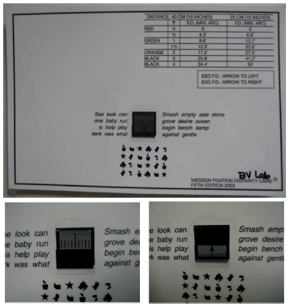
During testing with a Wesson fixation disparity card (top), monocular views through polarized glasses demonstrate how one eye sees the colored bars (bottom left) and the other eye sees the black arrow. The surrounding words provide fusion lock and control of accomodation. Each colored bar codes for a magnitude of fixation disparity indicated in the chart on the card.
Recommendations generally differ based on the type of heterophoria, namely:
Horizontal heterophoria. Prism is a viable option for treating patients with symptomatic binocular vision disorders. However, prisms may not work well for all patients equally. In a randomized clinical trial of the effectiveness of base-in prism reading glasses vs. placebo reading glasses for symptomatic convergence insufficiency in children, researchers found that base-in prism prescribed based on Sheards criterion was no better than placebo and thus concluded that BI prism was not an effective treatment for symptomatic convergence insufficiency in 9- to 17-year-old children.11 The effectiveness of BI prism for adults has not been studied.
Prism tends to be more effective for patients with divergence insufficiency, basic esophoria, and vertical heterophoria than for convergence insufficiency, basic exophoria, divergence excess and convergence excess.12,13 Therefore, prescribing universally for heterophorias based on any criterion may not alleviate your patients symptoms.
Nevertheless, when prism is the only feasible treatment for your symptomatic heterophoric patient, you may appreciate a mathematical review of two commonly used methods: These two methods require simple calculations based on the measured phoria and fusional vergence testing. (See Two Criteria for Determining Prism, above.)
Vertical heterophoria. Unlike horizontal heterophorias, there is more consensus about the best way to treat symptomatic vertical heterophorias. Vertical imbalances respond well to prism treatment.13-15
Two Criteria for Determining Prism
When prism is the only feasible treatment for your symptomatic heterophoric patients, you may apply these two methods:
~Sheard"s criterion. The compensating fusional reserve to blur point should be twice the amount of the heterophoria. The formula: Prism needed = 2/3(phoria) - 1/3(compensating fusional vergence).
So, if a patient has 6∆ exophoria and base-out (BO) to blur is 6∆, the prism needed would be 2/3(6) - 1/3(6), or 4 - 2. You would prescribe 2∆ base-in (BI), since deviation is exophoria.
~Percival Criterion. This formula states that the heterophoria should be in the middle third of the total range of fusional amplitude. The formula: Prism needed = 1/3(greater limit of BI or BO range) - 2/3(lesser limit of BI or BO range). Prism is only needed if this is a positive number.
So, for a patient who has 6∆ exophoria, BO rangers of 6/10/8 and BI ranges of 21/26/22, prism needed would equal 1/3(21) - 2/3(6), or 7 - 4. You would prescribe 3∆ BI since the deviation is exophoria.
Another example: A patient has 6∆ exophoria, BO ranges of 18/24/10 and BI ranges of 12/16/10, the amount of prism would equal 1/3(18) - 2/3(12), or 6 - 8, for -2∆. In this instance, no prism prescription is necessary.
Prescribing based on the vertical associated phoria is generally accepted as the best way to determine the proper amount of prism to treat symptomatic vertical heterophoria.13,16 This method also results in the least amount of prism needed to relieve symptoms.13 Associated phoria testing for determining prism to treat vertical heterophorias has become a standard method, going as far back as 1949 when Meredith Morgan Jr. reported 90% success in reducing symptoms by prescribing prism based on perceived vertical misalignments.16,17
While we have a widely accepted and accurate method of prescribing for vertical associated heterophorias, this method involves fixation disparity testing. Determining fixation disparity and associated phoria is relatively simple, quick and inexpensive. Several fixation disparity cards and slides are commercially available for less than $100.18 Or, consider polarized vectographic slides for distance testing, which cost about $300.18
The least expensive and simplest test often used is the Wesson fixation disparity card. The patient merely reports which colored line the arrow points at while wearing Polaroid glasses. Each colored line represents the fixation disparity measured in minutes of arc. The magnitude and direction of the prism required to neutralize or eliminate the fixation disparity is the associated phoria. This is accomplished by determining the amount of prism that makes the arrow point at the line indicating zero fixation disparity. The magnitude of prism that is the associated phoria is the prism amount to prescribe.
Prisms for Head Tilt
A 41-year-old white female presented with a longstanding history of lazy eye. She did not experience diplopia or asthenopia; however, she thought her lazy eye was getting worse because her children noticed her eye turn more frequently during the past year.
She presented with a head tilt to her right shoulder. When asked about this tilt, the patient denied having one until she pulled out three photo IDs from her wallet, all of which showed a noticeable right head tilt.
The Parks-Bielschowsky three-step test revealed a left hypertropia that increased in right gaze and left head tilt, indicating a problem with the left superior oblique. Further confirmation of the noncomitant deviation and muscle paresis was seen during the alternating cover test with prism neutralization in primary gaze. The deviation measured 25∆ when the prism was placed over her right eye (paretic left eye fixating). The deviation was only 10∆ when prism was placed over her left eye (non-paretic right eye fixating).
A trial frame of 5∆ BD over the paretic left eye, using a gross percentage criterion of one-half of the measured deviation, resulted in a visible straightening of the patients head. However, because the head tilt was longstanding and the patient reported no neck or back pains and no cosmetic concern about her head tilt, she chose not to have prism correction. Instead, she opted to try vision therapy to improve fusional vergence ranges. The patient had not started vision therapy at the time of this article.
Any time a patient presents with torticollis (unusual head posture), you must determine whether it is ocular torticollis, in which prism can be beneficial, or congenital torticollis, caused by a sternocleidomastoid muscle or vertebral malformation, in which prism is not beneficial.6 Ocular torticollis is a compensatory head posture caused by a binocular vision problem.19 If the patient straightens his or her head when you occlude one eye, you are dealing with ocular torticollis, not congenital.
Because the head position is compensatory in nature, cover testing with the patients head in a habitual posture may not reveal the offending deviation. Straighten the patients head when performing cover testing, and do not be surprised if the patient says his or her head does not feel straight when you do this. This is common when the condition is longstanding.
If the patients abnormal head posture is from a recent-onset, noncomitant deviation, it is critical to determine if he or she has a paretic strabismus or another potentially ominous underlying condition. Two clues that you are dealing with a paretic etiology: The deviation changes significantly when you place prism over one eye vs. the other during the alternating cover test, and prism differs when measured with the right eye vs. left eye is fixating. The deviation will be larger when the paretic eye is fixating (i.e., prism over the non-paretic eye during prism neutralization on the alternate cover test) and smaller when the nonparetic eye is fixating (i.e., prism over the paretic eye with prism neutralization on the alternate cover test).6
If you are dealing with a longstanding deviation or old paresis, the appropriate prism prescription can straighten the head significantly. Abnormal head positions are usually an indication of a noncomitant deviation. This is why you should perform the cover test in at least five gazes to determine type of noncomitancy: primary gaze, up gaze, down gaze, left gaze and right gaze. Sideways head turns compensate for horizontal deviations, such as with lateral rectus paresis, while head tilts are characteristic of oblique muscle problems, such as congenital or acquired superior oblique paresis. Also, vertical head tips are an indication of horizontal deviations that increase or decrease in up gaze or down gaze, known as A- or V-pattern deviations.
If the deviation is noncomitant, such as one caused by a paretic or A or V pattern deviation, yoked prisms that direct the patients eyes into the gaze with the smallest deviation will help straighten the head. If the torticollis is due to a vertical deviation, such as one caused by a superior oblique muscle paresis, vertical relieving prism rather than yoked prism is more appropriate. (See Postures and Treatments.)
Postures and Treatments Head Posture
Condition
Possible Cause(s)
Treatment
Left head turn
Right gaze preferred
Left lateral rectus (LR) paresis
Right medial rectus (MR) paresisYoked prism base left
Right head turn
Left gaze preferred
Right LR paresis
Left MR paresisYoked prism base right
Left head tilt
Right hyperdeviation
Right superior oblique (SO) paresis
Base-down over right eye if longstanding deviation
Right head tilt
Left hyperdeviation
Left SO paresis
Base-up over left eye if long-standing deviation
Head tip back
Downgaze preferred
V-pattern exotropia
A-pattern esotropiaBU yoked prism
Chin depressed
Upgaze preferred
A-pattern exotropia
V-pattern esotropiaBD yoked prism
Prism Adaptation
Patients with normal binocular vision for which prism is not required generally adapt to prism, as these patients tend not to be symptomatic.20,21 By contrast, patients who are symptomatic are less likely to adapt to prism, yet they are the most likely to benefit from it.22
Realize that some patients may have latent deviations that require more than one prism correction before they are completely compensated and stable. However, prescribe prism for your symptomatic patients with confidence that you will not induce any unwanted, irreversible prism adaptations with your treatment.
Dr. Tea is an assistant professor of optometry and chief of pediatrics and binocular vision at Nova Southeastern University College of Optometry in
1. Phillips PH. Treatment of diplopia. Semin Neurol 2007 Jul;27(3):288-98.
2. Aziz S, Cleary M, Stewart HK, Weir CR. Are orthoptic exercises an effective treatment for convergence and fusion deficiencies? Strabismus 2006 Dec;14(4):183-9.
3. OLeary CI, Evans BJ. Criteria for prescribing optometric interventions: literature review and practitioner survey. Ophthalmic Physiol Opt 2003 Sep;23(5):429-39.
4. Eustis HS, Mungan NK. Monovision for treatment of accommodative esotropia with a high AC/A ratio. J AAPOS 1999 Apr;3(2):87-90.
5. Caloroso EE, Rouse MW. Clinical Management of Strabismus.
6. von Noorden GK, Campos EC. Binocular Vision and Ocular Motility.
7. Steinman SB, Steinman BA, Garzia RP. Foundations of Binocular Vision. McGraw-Hill Companies; 2000:50-54.
8. Suchoff IB, Petito GT. The efficacy of visual therapy: accommodative disorders and non-strabismic anomalies of binocular vision. J Am Optom Assoc 1986 Feb;57(2):119-25.
9. Grisham JD. Visual therapy results for convergence insufficiency: a literature review. Am J Optom Physiol Opt 1988 Jun;65(6):448-54.
10. Griffin JR. Efficacy of vision therapy for nonstrabismic vergence anomalies. Am J Optom Physiol Opt 1987 Jun;64(6):11-14.
11. Scheiman M, Cotter S, Rouse M, et al.; Convergence Insufficiency Treatment Trial Study Group. Randomised clinical trial of the effectiveness of base-in prism reading glasses versus placebo reading glasses for symptomatic convergence insufficiency in children. Br J Ophthalmol 2005 Oct;89(10):1318-23.
12. Carter DB. Fixation disparity and heterophoria following prolonged wearing of prisms. Am J Optom Arch Am Acad Optom 1965 Mar;42:141-52.
13. Scheiman M, Wick B. Clinical Management of Binocular Vision;
14. London RF, Wick B. Vertical fixation disparity correction: effect on the horizontal forced-vergence fixation disparity curve. Am J Optom Physiol Opt 1987 Sep;64(9):653-6.
15. Sheedy JE, Saladin JJ. Association of symptoms with measures of oculomotor deficiencies. Am J Optom Physiol Opt 1978 Oct;55(10):670-6.
16. Cotter SA. Clinical Uses of Prism: A Spectrum of Applications.
17. Morgan MW. The Turville infinity binocular balance test. Am J Optom Arch Am Acad Optom 1949 Jun;26(6):231-9.
18. Bernell Catalog 2007;18-9.
19. Rubin SE, Wagner RS. Ocular torticollis. Surv Ophthalmol 1986 May-Jun;30(6):366-6.
20. Rutstein RP, Eskridge JB. Clinical evaluation of vertical fixation disparity. Part III. Adaptation to vertical prism. Am J Optom Physiol Opt 1985 Sep;62(9):585-90.
21. North R, Henson DB. Adaptation to prism-induced heterophoria in subjects with abnormal binocular vision or asthenopia. Am J Optom Physiol Opt 1981 Sep;58(9):746-52.
22. Schor CM. The relationship between fusional vergence eye movements and fixation disparity. Vision Res 1979;19(12):1359-67.
Prisms for Chin Tip
A 15-year-old Indian male presented accompanied by his mother, who said that her son always looks down and walks around with his head tilted back.
Cover testing revealed a V-pattern exophoria with insufficient compensating ranges in primary gaze. Deviation was noncomitant, measuring horizontally and 16s BI in upgaze, 10s BI in primary gaze, and 4s BI in downgaze. Cover testing in both eyes showed:
Through 2s BU yoked prism: 8s exophoria in primary gaze.
Through 3s BU yoked prism: 6s exophoria in primary gaze..
Through 4s BU yoked prism: 4s exophoria in primary gaze.
We allowed the patient to walk around with a trial frame of 4s BU yoked prism in each eye as well as his manifest refraction. He showed noticeable straightening of his head posture. We prescribed spectacles with yoked prism and presented the option of vision therapy to improve his compensating convergence ranges.
The new spectacles initially improved his head posture, but did not eliminate it completely. After 10 sessions of active vision therapy, both parents reported that they no longer observed the patient walking with a head tip. The parents opted to keep the current spectacles due to financial limitations, however agreed to try lenses without prism once their insurance approved a new spectacle prescription.
The patient has his head tipped back to compensate for V-pattern exophoria without and with yoked base-up prism glasses.
