 A 55-year-old black male presented with complaints of blurred vision at both distance and near. He reported that he only wore +2.00D over-the-counter reading glasses. The patient said that the glasses helped, but he still struggled to see.
His medical history was significant for HIV, which was diagnosed in 1992. Additionally, he reported hypertension and borderline high cholesterol. He did not know his CD4 number or his viral load, but indicated that he saw his doctor every three months. He took several medications, including Isentres (raltegravir, Merck), Prezista (darunavir, Janssen Therapeutics), Norvir (ritonavir, Abbott), PhenaTrim (weight loss supplement), acyclovir and lisinopril. He reported no known allergies.
On examination, his uncorrected visual acuity measured 20/200 O.D. and 20/100 O.S. With hyperopic correction, he was able to see 20/20 O.U. Confrontation visual fields were full to careful finger counting O.U. His pupils were equally round and reactive to light, with no afferent defect. Ocular motility testing was normal. The anterior segment examination was unremarkable. His intraocular pressure measured 13mm Hg O.U.
Dilated fundus exam showed clear vitreous without cells, as well as moderate-sized cups with good rim coloration and perfusion O.U. The vessels were of normal caliber and the maculae appeared normal. We noted multiple, peculiar lesions in each eye (figures 1 and 2). Additionally, we performed fundus autofluorescence (figures 3 and 4).
Take the Retina Quiz
1. What are the peculiar fundus lesions seen in both eyes?
a. Choroidal nevi.
b. Congenital hypertrophy of the retinal pigment epithelium (CHRPE).
c. Infiltrative metastatic carcinoma.
d. Primary malignant melanoma.
2. What is the significance of these lesions?
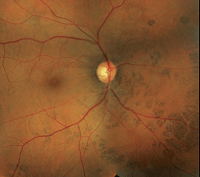
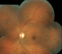
1, 2. Fundus photographs of our patient (O.D. left, O.S. right). Note the peculiar lesions scattered throughout the retinae.
a. There is no significance.
b. Diagnostic for metastatic disease.
c. Diagnostic for primary malignancy.
d. Marker for intestinal malignancy.
3. What is the correct diagnosis?
a. Multiple choroidal nevi.
b. Bear tracks.
c. Multiple CHRPE formation.
d. Gardner’s syndrome.
4. How should this patient be managed?
a. Observation.
b. Plaque radiotherapy.
c. Referral for intestinal malignancy.
d. Enucleation.
For answers, see below.
Discussion
The multiple lesions seen in both eyes represent a form of CHRPE. A typical CHRPE is a solitary, round, flat, well demarcated, hyperpigmented lesion that is present at birth.1,2 Its color can vary from dark brown or black to gray, and lacunae can be present within the lesion.
CHRPE are benign lesions that are usually discovered as an incidental finding during a routine eye exam. However, there are variations in how CHRPE may present.
One particularly interesting CHRPE variation presents as clusters or groups of lesions, as seen in our patient. In fact, if you use a little imagination, you almost get the impression that the pigmented lesions look like the paw prints of an animal that has been walking around the retina. For that reason, these lesions have been referred to as “bear tracks.”
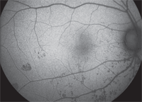
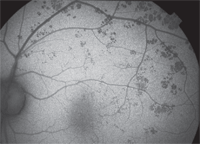
3, 4. Fundus autofluorescence reveals the presence of multiple pigmented lesions (O.D. left, O.S. right).
Bear tracks are congenital anomalies that are characterized by small, sharply circumscribed, variably sized, pigment spots. They are often grouped in one sector of the fundus. In our patient, they were predominantly located in the nasal retina, inferiorly in the right eye and superiorly in the left.
The bear tracks are significantly more apparent on fundus autofluorescence, where the hyperpigmentation causes evident blockages.
There has been some debate regarding the differences between CHRPE and bear tracks, because both presentations have a similar histopathology. In fact, it has been suggested that CHRPE and bear tracks are different expressions of the same condition.2 However, there are ultrastructural differences between the two entities. For instance, bear tracks are more elliptical in shape than CHRPE. In addition, when examined å electron microscopy, hypertrophy and hyperplasia of the RPE cells is not a significant feature in patients with bear tracks.2
The presence of multiple CHRPE lesions in both eyes should raise a concern for Gardner’s syndrome. Gardner’s syndrome, also known as familial colorectal polyposis, is an autosomal dominant disease that is characterized by adenomatous polyposis of the large and small intestines, hamartomas of the skeleton and various soft tissue tumefactions.
The risk for intestinal malignancy in adults with Gardner’s syndrome is virtually 100%.1,3 However, the shape of the CHRPE lesions seen in these patients is uniquely different than what is seen in patients with either classic CHRPE formation or bear tracks. More specifically, such lesions are not round like most CHRPE, but more oval––almost exhibiting a football shape.1
We were confident that our patient had nothing more than classic bear tracks. In addition, he had no history of familial adenomatous polyposis or any other colon problems. We explained the findings to our patient, gave him a prescription for glasses, and asked him to return annually.
1. Gass JD. Stereoscopic Atlas of Macular Disease: Diagnosis and Treatment, 4th ed. St. Louis: Mosby; 1997:805-9.
2. Regillo CD, Eagle RC Jr, Shields JA, et al. Histopathologic findings in congenital grouped pigmentation of the retina. Ophthalmology. 1993 Mar;100(3):400-5.
3. Buettner H. Congenital Hypertrophy of the Retinal Pigment Epithelium. In: Ryan SJ, Schachat AP, Murphy RP (eds). Retina, Vol. I––Medical Retina, 3rd ed. St. Louis: Mosby; 2000:634-9.
Answers to the Quiz
1. b
2. a
3. b
4. a
