 A 63-year-old white female presented with a chief complaint of blurry vision O.S. that was accompanied by floaters and flashes of light. She reported that the visual disturbances had progressively worsened over the past month.
Her medical history was significant for hypercholesterolemia as well as both cutaneous and lung malignancy. She underwent hysterectomy, tonsillectomy, resection of the cutaneous malignancy, and systemic chemotherapy and radiation. She was, however, free of metastatic disease at the time of initial presentation. Her current medications included simvastatin and Prilosec (omeprazole, AstraZeneca). She reported that she drinks socially and had never smoked.
On examination, her best-corrected visual acuity measured 20/25 O.D. and 20/100 O.S. Confrontation fields were full to careful finger counting in her right eye and constricted 360° in her left eye. On Amsler grid testing, she reported metamorphopsia in her left eye; however, her right eye was normal.
Extraocular motilities were full O.U. Pupils were equal, round and reactive to light, without evidence of afferent defect O.U. The anterior segment examination was significant for grade 1+ nuclear sclerotic cataracts in both eyes. Her intraocular pressure measured 14mm Hg O.U.
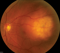
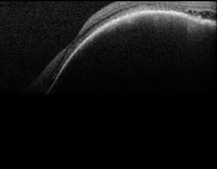
Dilated fundus examination of the right eye showed clear vitreous; a moderate-size cup with distinct margins; a flat macula; and a small (<0.5 disc diameter), flat, amelanotic lesion located superiorly in the periphery. The left eye showed a clear vitreous; a moderate-size cup with distinct margins; and a slightly elevated, creamy, yellow lesion (figure 1). Additionally, we performed an optical coherence tomography (OCT) scan (figure 2).
Take the Retina Quiz
1. What does the OCT scan reveal?
a. Posterior staphyloma.
b. Pigment epithelial detachment.
c. Choroidal mass.
d. Choroidal neovascular membrane (CNVM).
2. What test would be most helpful in establishing a diagnosis?
a. Visual fields.
b. Fluorescein angiography.
c. MRI.
d. Standardized ultrasound.
3. What is the correct diagnosis?
a. Metastatic carcinoma to the choroid.
b. Choroidal melanoma.
c. Choroidal hemangioma.
d. Posterior staphyloma.
4. How should this patient be managed?
a. Intravitreal anti-VEGF
injection.
b. Observation.
c. External-beam radiation therapy (EBRT).
d. Plaque radiotherapy.
|
| 1. Fundus photograph of the left eye shows a suspicious, elevated lesion located in the posterior pole.
|
5. What is the prognosis for this patient?
a. Excellent.
b. Guarded.
c. Too early to tell.
d. Poor.
For answers, see below.
Discussion
On clinical exam of the left eye, we observed a poorly circumscribed, creamy, yellow, elevated lesion with fluid that extended inferiorly. The elevation of the lesion was clearly evident on indirect binocular ophthalmoscopy. We noted that the lesion was intruding into the macular area, with extension of fluid toward the optic nerve.
In order to assess the area of fluid surrounding the lesion, we performed OCT. This showed a dome-shaped elevation of the RPE that originated from the choroid. Largely, this was due to a significant choroidal mass that was also causing a serous retinal detachment that extended inferiorly. Based on the patient’s history of lung carcinoma, we were highly suspicious that this finding was indicative of a metastatic lesion to the choroid.
Standardized echography was performed, which showed a dome-shaped lesion located posterior to the equator with dimensions of 12.5mm x 9.5mm and 3.6mm of elevation. It was irregularly structured with moderate internal reflectivity. The lesion was also shown to be mildly vascular. We confirmed the presence of the focal detachment at the inferior edge of the lesion on the B-scan. It is important to note that we uncovered no extraocular extension.
Based on the clinical appearance, you could potentially mistake this finding for an amelanotic choroidal melanoma. However, on diagnostic A-scan, a melanoma would have exhibited consistent low internal reflectivity. Additionally, we considered choroidal hemangioma in the differential diagnosis. These lesions show high internal reflectivity on the A-scan.
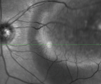
2. Confocal scanning laser image (top) and spectral-domain optical coherence tomography scan show interesting anatomical changes in the patient’s left eye.
Typically, metastatic choroidal lesions are a result of lung cancer in men and breast cancer in women. Additional, less likely sites of origin include cancer of the kidneys, thyroid, prostate and/or gastrointestinal tract.1 If the primary lesion site is unknown, biopsy by fine needle aspiration may be necessary.2
Although the mechanism is debatable, it is suspected that the vascularity of the choroid makes this location the most likely part of the eye for metastatic growth.3 It is extremely important to take a thorough history, because many patients may not disclose a history of cancer to their eye care providers. Once an adequate history has been taken, it is vital that these patients continue to be followed on dilated fundus examination, despite the state of their primary disease.
In our patient’s case, she had recently undergone a positron emission tomography (PET) scan and was told that she had no signs of active disease. However, we must keep in mind that the sensitivity of PET scans often cannot detect ocular lesions.
Once a diagnosis of metastatic carcinoma is made, the course of treatment should be expedited due to the potential for rapid lesion growth. Coordinated care among the eye care provider, oncologist and radiation therapist is critical for the success of treatment. The patient also will need to undergo a complete metastatic evaluation, including a PET scan.
In one report of 420 individuals with choroidal metastasis, 34% of the patients had no history of cancer at the time of diagnosis.1 Further, the tumors were unilateral in 320 patients and bilateral in 100 patients.1 Additionally, multifocal lesions were found in 20% of eyes, with a mean number of two lesions.1
In our patient, a second lesion thought to be an early “met” was seen superiorly in the right eye. Unfortunately, because it was located so far in the peripheral retina, we were unable to capture the entity on fundus photography.
In the past, once a metastatic lesion was detected in the eye, there was little consideration for treatment other than enucleation. Today, however, EBRT is the standard of care in bilateral cases. Plaque radiotherapy is occasionally used as well, especially in unilateral cases.4 Radiation therapy is especially important in cases where the lesion involves the macula or optic nerve.
In one published report, the researchers determined four indications for the use of EBRT in the treatment of metastatic breast carcinomas to the eye.4 These included secondary retinal detachment, measured decrease in visual acuity, threat for decreased visual acuity and rapidly enlarging tumor.4
Keep in mind that multidisciplinary, coordinated, individualized care often leads to the best outcomes for patients with choroidal metastasis.
Regarding our patient, we chose to perform EBRT due to the bilateral nature of the metastasis as well as the location of the lesion that extended into the macula and caused serous retinal detachment.
Thanks to Brigitte Keener, O.D., postgraduate optometry resident at Bascom Palmer Eye Institute in Miami, for contributing this case.
1. Shields CL, Shields JA, Gross NE, et al. Survey of 520 eyes with uveal metastases. Ophthalmology. 1997 Aug;104(8):1265-76.
2. Shields JA, Shields CL. Intraocular Tumors: A Text and Atlas. 4th ed. Philadelphia: W.B. Saunders Co; 1992:208-38.
3. Bloch RS, Gartner S. The incidence of ocular metastatic carcinoma. Arch Ophthalmol. 1971 Jun;85(6):673-5.
4. Mewis L, Young SE. Breast carcinoma metastatic to the choroid: analysis of 67 patients. Ophthalmology. 1982 Feb;89(2):147-51.
Answers to the Quiz
1. c
2. d
3. a
4. c
5. d
