 A 46-year-old Hispanic male presented with an eight-year history of reduced visual acuity in his right eye. He had seen several eye care providers over the years, but had always been confused as to why his vision was reduced. More recently, he began to notice blurry vision in his left eye.
Because this is his only functioning eye, he became extremely concerned and scheduled an evaluation. His medical history was significant for a brain aneurysm that required cranial surgery about five years ago.
On examination, his best-corrected visual acuity measured 20/400 O.D. and 20/30 O.S. Extraocular motility testing was normal. His confrontation visual fields were full to careful finger counting O.U.
His pupils were equally round and reactive to light and accommodation, with no evidence of afferent defect. Amsler grid testing revealed a dense central scotoma in the right eye and diffuse metamorphopsia in the left eye. The anterior segment was unremarkable.
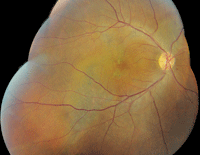
On dilated fundus examination, the patient’s vitreous appeared clear O.U. He had small cups with good rim coloration and perfusion in both eyes. Examination of the right macula showed nonspecific retinal pigment epithelium (RPE) mottling (
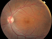
figure 1). We also noted RPE changes located temporally. In addition, we documented significant changes in the left macula (
figure 2).
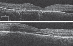
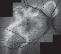
We obtained a spectral-domain optical coherence tomography (SD-OCT) scan of both maculae ( figures 3 and 4). Also, we performed fundus autofluorescence (FAF) on the patient’s right eye ( figure 5).
Take the Retina Quiz
1. What does the SD-OCT of the right eye reveal?
a. Occult neurosensory detachment and loss of the photoreceptor integrity layer (PIL).
b. Occult choroidal neovascularization (CNV).
c. Polypoidal choroidal lesions.
d. Occult persistent epithelial defect (PED).
2. What does the SD-OCT of the left eye reveal?


1, 2. Wide-field fundus photograph shows unusual changes in his right macula (left). Also, what do you notice about our patient’s left eye?
a. Shallow neurosensory detachment.
b. Shallow RPE detachment.
c. Occult CNV.
d. Polypoidal choroidal lesions.
3. What does the FAF scan of the patient’s right eye reveal?
a. Occult CNV.
b. Nonspecific atrophy of the RPE and photoreceptors.
c. Widespread RPE destruction and a trough line.
d. Loss of the RPE secondary to lipofuscin accumulation.
4. What is the most likely diagnosis?
a. Stargardt’s macular dystrophy.
b. Chronic idiopathic central serous choroidopathy (ICSC).
c. Occult CNV.
d. Polypoidal choroidal vasculopathy.
5. How do you believe the retinal specialist managed this patient?
a. Observation with protective eyewear.
b. Intravitreal anti-VEGF therapy.
c. Photodynamic therapy (PDT).
d. Laser treatment O.S.
For answers, see
below.
Discussion
Our patient has chronic ICSC in both eyes. The right eye exhibited nonspecific RPE changes in the macula. On clinical examination, there was no evidence of subretinal fluid; however, the SD-OCT clearly shows shallow puddles of fluid scattered throughout the macula. Interestingly, the fovea seems to be spared. But, that probably wasn’t always the case because the SD-OCT scan reveals extensive loss of the PIL, which partially explains the 20/400 visual acuity.

|
| 3,4. Spectral-domain optical coherence tomography scan revealed some interesting findings (O.D. top, O.S. bottom).
|
The larger area of RPE mottling located temporal to the right macula that was seen on wide-field color photography is of particular interest. The presentation appears as a linear area of RPE mottling that extends inferiorly. This represents a trough line that developed as a result of fluid leakage from the serous detachment.
The FAF scan of the right eye is also quite remarkable, because it clearly illustrates the extent of RPE degeneration located throughout the macula and posterior pole in a radiating, star-like pattern. The trough line that is seen extending inferiorly also is better appreciated on FAF.
FAF is a noninvasive photographic method of documenting pathological changes within the retina and the RPE. In patients with AMD, FAF commonly is used to evaluate the effects of lipofuscin accumulation within the RPE. In this case, the use of both FAF and SD-OCT helps us understand what happened in the patient’s right eye, uncovering the presence of persistent fluid, damage and destruction to the RPE and photoreceptors.
The clinical examination of his left macula revealed a shallow neurosensory detachment, which we confirmed on SD-OCT. Further, we noted fine, yellow precipitates located on the inner retinal surface. Fortunately, his visual acuity remained widely unaffected O.S.
Most cases of ICSC resolve naturally without any treatment. However, recurrences develop in approximately 33% to 50% cases within one year.1 It is interesting to note that 10% of patients will experience three or more episodes or ICSC.1 Clearly, our patient falls into this category.

5. A fundus autofluorescence scan of the patient’s right eye. How do you explain these findings?
Given the history of progressive vision loss in his right eye, as well as the presence of a shallow serous detachment in his left eye, we referred our patient to a retinal specialist. The patient was seen within a two-week period. The retinal specialist confirmed the findings that we saw in the patient’s right eye. Interestingly, however, the serous detachment in his left eye had completely resolved at the time of his appointment.
Nonetheless, the retinal specialist elected to follow the patient, without treatment, and instructed him to return in six weeks.
1. Gass JD. Stereoscopic atlas of macular disease: Diagnosis and treatment. 4th ed. St. Louis: Mosby; 1997:52-70.
Answers
1. a
2. a
3. c
4. b
5. a

