 A 35-year-old black female presented for an annual eye examination. The patient reported that she experienced transient blurry vision at distance and near, which lasted for a few minutes at a time. Her last eye exam was four years ago, and she did not wear glasses. Her medical history was unremarkable.
On examination, her entering visual acuity measured 20/20 O.U. Confrontation visual fields were full to careful finger counting O.U. Her pupils were equally round and reactive, without evidence of afferent defect. Extraocular motility testing was normal. The anterior segment examination of both eyes was unremarkable. Intraocular pressure measured 14mm Hg O.U.
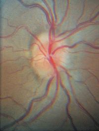
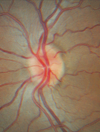
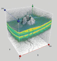
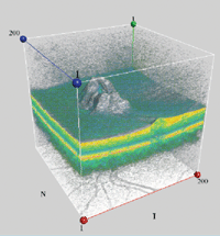
On dilated fundus examination, the vitreous was clear in both eyes (figures 1 and 2). The macula, vessels and periphery were normal. Additionally, we obtained a spectral domain optical coherence tomography (SD-OCT) scan of both eyes (figures 3 and 4).
Take the Retina Quiz
1. How would you characterize the findings in her optic nerves?
a. Normal.
b. Pseudopapilledema.
c. Generalized disc swelling.
d. Hypoplastic.
2. What additional testing is necessary?


1, 2. Fundus photographs of our patient (O.D. left, O.S. right). Look closely at both optic nerves. What do you notice?
a. No additional testing is necessary.
b. Automated visual fields.
c. MRI and lumbar puncture (LP).
d. Both b and c.
3. Based on the clinical findings and SD-OCT scan, what is the most likely diagnosis?
a. Optic nerve head drusen.
b. Papilledema.
c. Optic nerve hypoplasia.
d. Idiopathic intracranial hypertension (IIH).
4. How should this patient be managed?
a. Observation.
b. Periodic visual fields.
c. Refer for possible MRI and LP.
d. Optic nerve sheath fenestration.
For answers, see below.
Discussion
Our patient has bilateral optic nerve swelling (O.D. > O.S.). On clinical examination, the optic nerve clearly was elevated and the margins were blurred. We confirmed this finding on SD-OCT, where the retinal nerve fiber layer was thickened beyond what is observed in the normative database. This was highly evident on the 3D topographic OCT image. We documented no flame hemorrhages or pattern lines, which are often present in severe cases of optic nerve edema.
So, what’s wrong with our patient? After careful evaluation, we determined that she had IIH. Based on the patent history provided at this visit, it would be almost impossible to make a diagnosis of IIH in the absence of an MRI and an LP. Fortunately, we had a previous history to rely on.
At her last eye examination four years earlier, she was under the care of a neuro-ophthalmologist for IIH. At that time, she presented with ringing tinnitus and swollen optic nerves. She had the typical physical profile of a patient with IIH, with a height of 5 feet 6 inches and a weight of 180 pounds.
At that visit, an MRI and LP were ordered. The MRI was normal. However, the LP revealed opening pressures of 330mm H2O (the normal range is 70mm H2O to 180mm H2O). Additionally, we obtained serial visual fields, which showed generalized constriction with limited reliability.


3, 4. Three-dimensional macular cube scans of our patient’s optic nerves on spectral domain optical coherence tomography (O.D. left, O.S. right).
Previously, we had prescribed 1,000mg of acetazolamide per day to lower her intracranial pressure. She also received counseling on the health benefits of weight loss. But subsequently, she was lost to follow-up.
IIH develops secondary to chronically elevated intracranial pressure. Over the years, increased intracranial pressure has been described by several different terms––most commonly pseudotumor cerebri and benign intracranial hypertension (BIH).1 Today, IIH is the preferred term.
The most significant neurologic complication caused by IIH is papilledema. If untreated, papilledema may lead to progressive optic atrophy and blindness. Other neurologc symptoms of IIP include severe headaches, ringing in the ears, blurred vision and diplopia (which may result from a cranial VI palsy).1
IIH predominately affects obese women of childbearing age.1 There is no known etiology; however, there are several risk factors for the disease, including exposure to or withdrawal from certain exogenous substances (e.g., pharmaceutical agents, such as tetracycline), a wide variety of systemic diseases (e.g., Lyme disease, anemia, lupus and sarcoidosis), numerous endocrine or metabolic disorders, and disruption of cerebral venous flow.1,2
The treatment for IIH is multifaceted. Weight loss is considered to be the best long-term management objective for this condition, but often proves to be difficult. But, even slight weight reductions can make a significant difference. In one study, for example, just a 5% to 10% decrease in overall body mass yielded a reduction in intracranial pressure and facilitated papilledema resolution.2,3 Nonetheless, referral to a dietitian may be appropriate to help formalize this process.
The most effective pharmacologic treatment for IIH is acetazolamide. The initial dosage is 500mg to 1,000mg/day (e.g., 500mg Diamox Sequels [acetazolamide, Duramed Pharmaceuticals] b.i.d.).1 Unfortunately, many patients become intolerant due to the side effects of the medication, including paresthesias in the extremities, fatigue, metallic taste from carbonated beverages, and decreased libido.
For patients who report visual function decline, surgical management may be warranted. Appropriate interventions include optic nerve sheath fenestration, lumboperitoneal/ventriculoperitoneal shunt implantation to divert cerebrospinal fluid, and venous sinus stenting.1,2
Our patient seemed to be doing well, despite not undergoing an examination in four years. Even though there was generalized swelling of both optic nerves, their color still appeared healthy. Color vision testing also was normal, and visual field results were stable compared with those obtained on previous evaluations.
The patient was referred to a neuro-ophthalmologist a few weeks later, and acetazolamide was restarted. We instructed her to return in four months for follow-up.
1. Friedman DI. Papilledema. In: Miller NR, Newman NJ, Biousse V, Kerrison JB (eds). Walsh & Hoyt’s Clinical Neuro-Ophthalmology. 6th ed. Vol I. Philadelphia: Lippincott Williams & Wilkins; 2005, 237-91.
2. Gans MS. Idiopathic Intracranial Hypertension. Medscape. Available at:
http://emedicine.medscape.com/article/1214410-overview. Accessed November 12, 2012.
3. Kupersmith MJ, Gamell L, Turbin R, et al. Effects of weight loss on the course of idiopathic intracranial hypertension in women. Neurology. 1998 Apr;50(4):1094-8.
Answers
1. c
2. b
3. d
4. b

