An active 31-year-old female walks into your office with an unremarkable medical history; she’s a non-smoker who falls within a healthy weight range. She complains that recently she has seen a small, dark spot in her vision in one eye for a short period of time and has had a history of ophthalmic migraines in the past.
It would be tempting to assume that the “dark spot” she saw was just a poorly described ophthalmic migraine—so tempting to assume, that even I took the bait and diagnosed it as just that. That young, healthy 31-year-old woman was me. And it wasn’t an ophthalmic migraine I was experiencing, it was a transient ischemic attack (TIA), also known as a mini stroke.
Although my experience was terrifying and unsettling, it gave me a better understanding of what it feels like to be on the other side of the patient-doctor interaction and has allowed me to improve communication and patient care in my practice.
How it Happened
I was at home making my kids their lunches when I saw a small distortion in my vision and thought, “Oh boy, here we go, another ophthalmic migraine.” I cursed the fact that, for the next 20 minutes, my vision in that eye would soon become a swirling blur of zigzags and zebra stripes. The only weird thing was that the visual defect never radiated outward like the ophthalmic migraines I had experienced in the past. Its lack of expansion really didn’t strike me as odd at the time, so I just continued on and finished up lunch.
About 10 minutes later, I was reading my son a book when my ability to read was suddenly turned off like a light switch. I was completely and instantaneously illiterate. I could see letters on the page. I could see that they were grouped into words. But I had no idea what those words were—it was as if they were written in a foreign language.
Frightened, I grabbed the phone and called my husband and other family members who lived nearby. They were alarmed, but assumed that I was dehydrated and encouraged me to drink fluids and eat something. I knew it was more than that. My family and I decided that someone needed to take me to the doctor or the ER. While I was grabbing my purse and cell phone, I became so nauseated and dizzy that I almost fell over. At this point, we called 9-1-1.
I lay on the bench in my hallway, waiting for the ambulance to arrive, while the doctor in me still tried to self-diagnose the situation. It has to be poisoning, I thought. I must have touched some household chemical underneath the sink, and then accidentally ingested it somehow. Soon, I began to slur my speech; then, as my right arm and the right side of my face went numb, I sadly congratulated myself on finally realizing the correct diagnosis. Stroke.
Failure to Communicate
In the ambulance and ER, my speech improved as they gave me oxygen. The slurring was minimal; however, I could not form the right words. I tried to explain the initial dark spot in my vision to the doctors , but I could not say the word “vision”—I kept saying “version.” In my mind, I was cognitively all there. I was stunned and astonished to hear the sound of my own voice, disembodied from me, saying the wrong word. I repeated the word several times, but it came out wrong every time. I couldn’t believe what I was hearing from my own mouth!
I thought maybe I could communicate with them better by writing so I motioned for the nurse to give me paper and a pen, thinking I would give them a hand-written account of what had happened. But each and every time I went to say the words “hand written,” it came out as “hard,” and she couldn’t understand what I was requesting. I was flabbergasted. I felt trapped in my own mind, unable to communicate. I saw the looks on the faces of the nurses and doctors and knew that they had given up on me being able to tell them what had happened. It was a horrible feeling. It has made me more sympathetic to patients in my chair who have a tough time communicating with me as their doctor, and it has given me more patience in listening to what it is they’re trying to say.
Over the next few hours in the ER, I must have had every test in the book—including blood work, chest X-rays, a CT scan, carotid Doppler ultrasounds, a magnetic resonance angiogram and an MRI of my head, neurological testing and an electrocardiogram. Absolutely everything came back normal. The doctors told me the good news was that I was definitely a very healthy young woman; the bad news was that I had experienced a TIA.
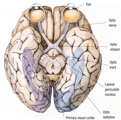
A TIA often affects the anatomy of the visual pathway, frequently manifesting as severe monocular vision loss or blindness that occurs suddenly and resolves in less than five minutes.5
With an ischemic stroke, a blood clot blocks an artery in the brain and stays there. With a hemorrhagic stroke, a blood vessel bursts in the brain. But with a TIA or mini-stroke, a blood clot blocks an artery in the brain temporarily and then moves on.
Because my cholesterol levels were normal and I had no blood clotting disorders, they believed a positional blood clot had caused the TIA. They suspected the blood clot had formed in my leg because it wasn’t getting proper circulation due to the way I was sitting on the floor while I was reading the book. They imagined the blood clot must have been smaller than the head of a pin, and that it traveled to my brain, where it stayed for the next several hours. Eventually, it dislodged itself and went on its way.
The doctors asked me questions about my heart health, and if I had been born premature. I was delivered at full term, and although I had a family history of heart disease, I never experienced any heart problems or murmurs myself. They told me that we all get tiny clots and debris in our blood at times, and that usually our lungs filter them out. But, sometimes, an otherwise healthy person can suffer a TIA because a tiny blood clot goes straight to the brain, bypassing the normal circulation route through the heart and filtration through the lungs.
The Culprit
In a healthy heart, normal blood flow moves from the body to the heart, through the lungs back to heart, and then finally gets redistributed through the body. After circulating through the body, deoxygenated blood returns to the heart and enters the right atrium. Then, it goes through the tricuspid valve to the right ventricle, and exits the heart from the pulmonary valve.
Next, the pulmonary arteries carry the blood to the lungs to get fresh oxygen. The re-oxygenated blood travels back to the heart via the pulmonary veins and enters the left atrium; then, it passes through the mitral valve to the left ventricle. The left ventricle pumps the oxygenated blood to the aorta and other arteries, where it gets redistributed to the body and brain.1
A patent foramen ovale (PFO) is a hole in the atrial septum, the wall that divides the left and right atria (upper chambers of the heart). Everyone has this hole while in the womb, but usually the hole closes shortly after birth. However, in 20% of people, it does not.2 Tiny blood clots formed in the body can use this congenital heart defect in the atrial-septal wall as a door to return to the heart and go straight to the brain, instead of going through the normal filtration and re-oxygenation system of the lungs. Preemies are sometimes born with PFOs, which is why neonatal intensive care units screen for them, but a full-term baby (the average person) never gets screened for a PFO.
A transesophageal echocardiogram (TEE) is used to detect a PFO in adults, using a transducer (sort of like a microphone) to send sound waves through the heart. These waves bounce off different structures in the heart, and help to assess the heart’s function and check for structural abnormalities. The transducer is fed down the esophagus, while the patient is under general anesthesia. Going through the esophagus yields a clearer image of the heart from its back side because the sound waves do not have to travel through the skin, bones and muscle like they would if the transducer were placed over the heart on the outside of the chest.
When all of my other tests came back absolutely normal, the doctors recommended a TEE, so I fasted and underwent the procedure the next morning. Lo and behold, there it was—I indeed had a PFO.
Once a PFO is diagnosed in a patient who has had a TIA, the doctor may prescribe blood-thinners if they feel that line of treatment may help to prevent another clot from forming and causing a repeat TIA or stroke.3 Another option is to have an umbrella-like device implanted, which closes the holes permanently once it’s positioned in the atrial-septal wall and opened. It’s not open-heart surgery; the entire procedure is done through a catheter fed through a vein in the leg. Trials are still ongoing to determine whether anti-coagulant therapy and PFO closure surgery are equally as effective when it comes to reducing the likelihood of a second TIA or stroke. The decision ultimately lies in the hands of the doctor and the patient.
Closing the Door
Signs and Symptoms4
It’s important to know and watch out for the signs and symptoms of a stroke. Seek medical attention immediately if you have any of the following:
• SUDDEN numbness or weakness of face, arm or leg—especially on one side of the body.
• SUDDEN confusion, trouble speaking or understanding.
• SUDDEN trouble seeing in one or both eyes.
• SUDDEN trouble walking, dizziness, loss of balance or coordination.
• SUDDEN severe headache with no known cause.
Forty-two days after my TIA, I had PFO closure heart surgery. I find comfort in the fact that this pathway is now closed, and I am hoping to live the rest of my life stroke-free. I am extremely lucky that I haven’t had any residual effects from the mini-stroke and that I survived. It is impossible to know if what you are experiencing is a TIA, ischemic stroke or hemorrhagic stroke, and proper medical intervention is absolutely necessary to protect your health and your life. (See “Signs and Symptoms,” right.)
To this day, I still shake my head at the fact that I misdiagnosed myself! That little black spot in my vision. Although I disregarded it at first, at least I had enough sense to be on high alert for any other peculiar signs or symptoms that popped up. The doctors say it was good that I acted fast and did not ignore my other symptoms. I got oxygen quickly from the ambulance because of calling 9-1-1 right away—and, in a stroke, that matters.
Having a TIA is a potential warning sign of a future stroke or some serious underlying issues. That is why I feel it is so important for us as optometrists to be on the lookout for weird symptoms and stories from our patients, even in those who are young and seem completely healthy.
We are trained that “when you hear hoof beats, think horses, not zebras,” but as a zebra, I can tell you from personal experience, sometimes the most likely diagnosis is not always the correct one.
Dr. Murphy practices full-scope optometry in Holbrook, N.Y.
1. American Heart Association. How the healthy heart works. Available at:
www.heart.org (accessed February 7, 2012).
2. PFO Research Foundation. PFO overview. Available at:
http://pforesearch.org/about/pfo-overview (accessed February 21, 2012).
3. National Stroke Association. What is TIA? Available at:
www.stroke.org (accessed February 21, 2012).
4. National Stroke Association. Warning signs of stroke. Available at:
www.stroke.org (accessed February 7, 2012).
5. Hubel DH. Eye, Brain and Vision. New York: Scientific American Literature, 1988.
