Optometrists play a key role in diagnosing and managing guttata and Fuchs’ dystrophy. Previously, when conventional treatments (e.g., sodium chloride drops, aggressive ocular surface disease therapy, heat therapy) couldn’t adequately manage the condition, our only surgical option was a penetrating keratoplasty.
Fortunately, corneal transplant surgery has been evolving continuously from a full-thickness procedure to several partial-thickness surgeries—chief among them Descemet’s stripping automated endothelial keratoplasty (DSAEK). It has revolutionized surgical intervention for endothelial dysfunction because it leaves the anterior cornea structurally intact, allows faster healing and VA recovery, reduces graft rejection risk, lessens post-op astigmatism and allows for stronger wound integrity.
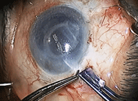
DSAEK is a sutureless transplant of the posterior cornea that replaces only the diseased portion with a donor graft. This 45-minute procedure can be performed alone or in combination with cataract surgery.
The endothelium is stripped and scored around the peripheral stroma to promote adhesion of the donor tissue. Next, the surgeon prepares the donor tissue, which is measured, centered and trephinated to ensure proper sizing. The donor graft is created by either manual dissection using a microkeratome (DSEK) or automated dissection with a femtosecond laser (DSAEK). The graft—typically about 150µm thick—consists of posterior stroma, Descemet’s membrane and corneal endothelium.
Viscoelastic is placed on the donor endothelium to protect it during insertion. The donor tissue is folded and inserted using specialty forceps to minimize insult to the delicate tissue. The surgeon centers the graft with an injection of balanced saline, then fills the entire anterior chamber with sterile air to aid graft attachment. Additional surgical measures taken include peripheral iridectomies and properly sized air tamponades to prevent pupillary block glaucoma. Once the graft is stabilized, the wound is sealed and closed, and a pressure patch is secured.
Multimedia
Go to
http://www.revoptom.com/multimedia/ to watch how Descemet’s stripping automated endothelial keratoplasty is done.
On the one-day post-op exam, vision is approximately <20/200. IOP is measured and the peripheral iridectomy is checked for patency. Mild to moderate corneal edema may be present. The anterior chamber should be filled with 30% air. Patients are encouraged to remain supine for several days to promote graft adherence. The follow-up schedule typically is one day, one week, then monthly to ensure proper healing and recovery. At six months, the majority of patients see >20/40.
DSAEK has been a promising development in corneal surgery, and the procedure is constantly evolving. Some surgeons are starting to use thinner grafts (Descemet’s membrane endothelial keratoplasty, or DMEK), which improve visual outcomes but require a more technically challenging procedure. In the meantime, DSAEK is the current standard for endothelial disease. As optometrists, it is important for us to understand the latest surgical procedures so we can educate and treat our patients accordingly.
