Many dystrophies can affect the endothelial cell layer of the cornea. Given the limited magnification available on a slit lamp, it is difficult to visualize changes in the endothelium with much precision, especially during the early stages of dystrophic endothelial diseases. Many times, these types of dystrophies go undiagnosed until the damage has progressed.
To that end, non-contact specular microscopy allows optometrists to visualize and diagnose dystrophies earlier and more accurately.1 Having the ability to perform endothelial cell counts noninvasively allows eye care practitioners to provide quicker, more accurate diagnosis and treatment.
Specular microscopy also offers valuable insight when making decisions that range from contact lens selection to surgical referral. While the hefty price tag on this technology may deter some practices, others will find it a worthwhile investment that pays off not just in better patient care but also in reimbursement dollars.
How It Works
 At Nittany Eye Associates, we use a CellChek XL clinical specular microscope system (Konan Medical), which allows us to visualize the corneal endothelium at the cellular level in vivo. The endothelium, the most posterior layer of the cornea, is a monolayer of approximately 350,000 to 500,000 highly specialized cells.2
As these cells do not have the ability to replicate, humans are born with the maximum number of endothelial cells they will ever have. Histologically, these cells initially have a hexagonal shape. As endothelial cells die, neighboring cells enlarge to cover the empty space once occupied by the cell. This, in turn, causes the remaining cells to lose their hexagonal shape.
The specular microscope projects light onto the cornea, and then captures the image that is reflected from the optical interface between the corneal endothelium and the aqueous humor.
CellChek XL quantitatively analyzes this information and generates four numeric indices: cell density (CD), coefficient of variation (CV), percentage of hexagonal cells (HEX) and number of cells used to calculate the results (NUM). The first three indices are useful for measuring endothelial cell death.
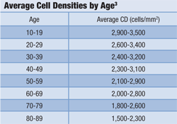
- CD is a measurement of cell density in mm2 and decreases with age. (See “Average Cell Densities by Age.") A low CD value for a particular age may indicate that the endothelium is depleting faster than normal.
- CV represents the coefficient, or degree, of variation in the sizes of the endothelial cells (polymegethism). By measuring the variation in size between endothelial cells, the system can measure how much cell loss is occurring. A CV less than 40 is normal.
- HEX indicates the variability in hexagonal cell shape over time. Hexagonality above 50% is suggested to be normal.3
Clinical Benefits in Practice
The ability to properly diagnose and treat corneal endothelial dystrophies allows optometrists to provide a high level of care. Knowing the integrity of the endothelium is another useful tool in a cornea and contact lens practice. In addition, it can explain seemingly idiopathic decreases in vision by revealing subclinical corneal issues.
Taking an endothelial cell count can be helpful before referring patients for cataract or refractive surgery so that any potential endothelial dysfunction or thinning is recognized before they have the surgical intervention. Pre-existing corneal conditions, such as Fuchs’ dystrophy, dry eye, epithelial basement membrane dystrophy and Salzmann’s nodular degeneration, can decrease the potential for positive surgical outcomes.4 This information provides valuable insight to both the referring optometrist and the surgeon.
At Nittany Eye Associates, the cataract surgeon may lower the phacoemulsification energy in an attempt to preserve endothelial function in patients with a mildly compromised endothelium as determined by specular microscopy. One frequently-touted benefit of the new “femto-phaco” cataract surgery is its potential to be less traumatic to the endothelium by reducing the phaco energy used during the procedure; prescreening with specular microscopy could identify patients better suited to this approach than conventional cataract surgery.
An endothelial cell count also can play an important role in fitting a patient with the right type of contact lens. For example, overnight wear may work successfully in someone with a properly functioning endothelium, but it may cause a variety of issues in a patient with inadequate endothelial function.
With the evolution of lens materials and wearing schedules, there are many options in the optometrist’s arsenal. Having this information available before fitting patients in contact lenses can allow the eye care practitioner to make an informed decision when choosing the appropriate contact lens.
In our practice, specular microscopy has served as a helpful aid in diagnosis of endothelial dysfunction and other underlying corneal issues, as illustrated in the following case examples.
Case Examples
Fuchs’ Endothelial Dystrophy
A 54-year-old white female presented with a chief complaint of reduced distance and near vision in her left eye. BCVA was 20/20 O.D. and 20/40 O.S.
External testing was unremarkable. Slit lamp examination was remarkable for a filtering bleb and laser peripheral iridotomy in the left eye. There was temporal corneal scarring in the left eye as well as striae in the corneal stroma. General corneal haze was noted in both eyes, but was more significant in the left. Her dilated fundus examination was unremarkable.
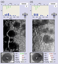
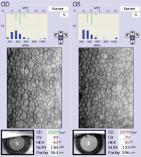
1. Fuchs’ endothelial dystrophy—note the significantly increased CV and decreased HEX in the left eye.
Specular microscopy of the left eye shows a significantly increased CV as well as a significantly decreased HEX (figure 1). This information reveals cell loss and subsequent morphing of surrounding cells to fill in the compromised endothelium.
Her pachymetry shows thickened corneas, measured at 630µm O.D. and 601µm O.S., further confirming the endothelial dysfunction. In addition to grade three guttata, coalescing guttatae are noted in the cell count image. Due to these signs—particularly the guttata and abnormal CV values—we diagnosed the patient with Fuchs’ endothelial dystrophy.
Fuchs’ is a dysfunction of the cornea’s endothelial layer that typically presents between the fifth and sixth decade of life, and affects women more than men.5 Be sure to question patients about their family history, as Fuchs’ is inherited in an autosomal dominant fashion.6 Treatment of Fuchs’ typically involves hyperosmotic agents to pull excess fluid out of the cornea to reduce edema. IOP-lowering medications, such as topical prostaglandins or beta-blockers, are also used in patients with increased IOP.
Limit use of medications preserved with benzalkonium chloride, as chronic exposure may be toxic to endothelial cells.7 In advanced cases where topical agents cannot combat corneal edema, corneal grafts are inserted. Currently, the surgical treatment of choice is Descemet’s membrane stripping automated endothelial keratoplasty (DSAEK).8
Chandler’s Syndrome
A 54-year-old white male presented with a chief complaint of blur at near in the left eye. His BCVA was 20/20 O.D. and 20/40 O.S. External testing was unremarkable. Slit lamp examination revealed a peripheral iridotomy and a filtering bleb in the patient’s left eye.
The left cornea also showed corneal scarring temporally and a diffuse haze noted throughout the cornea, including centrally. The dilated fundus examination was unremarkable.
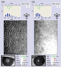
2. Endothelial cell count indicative of Chandler’s syndrome, O.S.
Specular microscopy data from the left eye shows normal cell counts, but abnormal CV and HEX indicates pleomorphism is occurring (figure 2). The left endothelial cell layer clearly shows blurred cell margins. This patient has a common presentation of Chandler’s syndrome, a disorder characterized by abnormal proliferation of the cornea’s endothelial layer.
The excess endothelial cells adhere to the iris and lodge themselves in the trabecular meshwork. This results in distortion of the iris, increased IOP and corneal edema. Chandler’s syndrome frequently presents in young adulthood or middle age and often is found unilaterally with the uninvolved eye manifesting only subclinical signs.9
This holds true in the patient’s scan. The right eye (the uninvolved eye) clearly shows a more uniform and functioning endothelium. In addition, the pachymetry readings show the left cornea is thicker than the right eye, indicating greater edema O.S. Specular microscopy allowed us to confirm this diagnosis, and is a valuable tool in identifying the clinical signs of Chandler’s syndrome.
Treatment of Chandler’s syndrome often centers on controlling the corneal edema and managing increased IOP. Corneal edema is treated with hyperosmotic solutions and ointments in the early stages. In advanced cases, a corneal transplant is warranted. Increased IOP is managed with glaucoma medications.
Agents that decrease aqueous production, such as beta-blockers, yield better results than those that work by increasing aqueous outflow. This is because the endothelial pumps are not working at sufficient capacity to successfully manage the existing aqueous.8
Contact Lens Intolerance
A 31-year-old white male had a history of contact lens intolerance. He reported decreasing vision throughout the day. His BCVA was 20/20 O.D. and 20/20 O.S. His contact lens history was lengthy and included many different materials as well as various replacement schedules—all with no relief of symptoms.
His endothelial cell count in a 26 Dk daily lens was normal, with a decreased CV and HEX in the right eye (figure 3). While CD is normal, the decreased CV and HEX indicate cell loss.
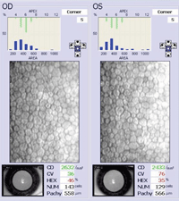
3, 4. Endothelial cell count of patient with contact lens intolerance in a 26 Dk daily lens (top). Endothelial cell count of same patient with contact lens intolerance in a 55 Dk daily lens.
The left eye demonstrates decreased CD, CV and HEX, which suggests a dysfunctional endothelial cell layer and explains the patient’s contact lens intolerance and decrease in vision throughout the day. The patient was instructed to discontinue contact lens wear for two to three weeks. After this time, the patient was to resume contact lens wear with a 55 Dk daily lens to increase oxygen transmission to the ocular surface.
The endothelial cell count was repeated after approximately three months (figure 4). The right eye showed an improved CV, and there was also an increase in CD of the left eye. While endothelial cells do not regenerate throughout a person’s lifetime, the increase in CD may be a reflection of a healthier endothelium and a more reliable reading.
The pachymetry readings were also decreased compared to the previous cell count, which indicates resolution of prior corneal edema. This cell count suggested to us that increasing the oxygen permeability of the patient’s contact lenses would allow the endothelium to function at a higher level, thus permitting longer wear time and improved comfort.
With advances in high-Dk lens materials and FDA approval of overnight contact lens wear, assessing corneal integrity is more important than ever. Being able to analyze important endothelial characteristics provides eye care practitioners with another tool to evaluate corneal health. It also allows practitioners to advise against overnight wear or lower Dk lenses in those patients who have dysfunctional endothelial cells.
The ability to assess the endothelial cell layer has allowed clinicians in our practice to look at additional facets of the patient’s corneal health that can help to explain why a seemingly healthy person is unable to comfortably and successfully wear contact lenses.
In our practice, using specular microscopy has helped us to better predict which type of lens (i.e., high Dk, daily replacement) will work for the patient and reduce frustration.
Does It Pay Off?
Although the initial investment in a specular microscope may cost your practice anywhere from $25,000 to $30,000 per unit, manufacturers offer monthly payment plans and leasing options to make the cost more feasible. Reimbursement also can help you get a reasonable return on your investment.
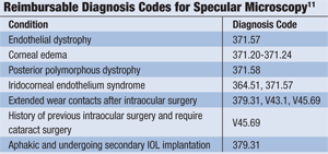
Insurance companies will reimburse for specular microscopy when medically necessary, which can provide a significant addition to practice revenue. The current CPT code for specular microscopy is 92286 (special anterior segment photography).
As it is a bilateral code, the reimbursement is the same whether specular microscopy is performed on one or both eyes.11 Medicare currently reimburses the procedure for a wide variety of diagnoses. (See “Reimbursable Diagnosis Codes for Specular Microscopy.")
Endothelial cell photography is a covered procedure under Medicare when reasonable and necessary for patients who meet one or more of the following medical criteria:12
- slit lamp evidence of endothelial dystrophy (corneal guttata).
- slit lamp evidence of corneal edema (unilateral or bilateral).
- about to undergo a secondary intraocular lens implantation.
- had previous intraocular surgery and require cataract surgery.
- about to undergo a surgical procedure associated with a higher risk to the corneal endothelium (i.e., phacoemulsification or refractive surgery, with evidence of posterior polymorphous dystrophy of the cornea or irido-corneal-endothelium syndrome).
- about to be fitted with extended wear contact lenses after intraocular surgery.
It is important to know Medicare’s coverage policy for specular microscopy before cataract surgery.11 Simply stated, if performing cataract surgery in an individual for the first time whose only visual issue is cataracts, specular microscopy is covered as part of the presurgical work-up. The patient’s record should contain: an order for the test, an endothelial image, test reliability, findings, diagnosis and doctor’s signature.11
Medicare currently reimburses about $85, while other third-party payers reimburse $120.12 These rates are national averages and vary throughout the country. The break-even point is between 250 and 300 scans.
In one year, we performed more than 100 scans at Nittany Eye Associates. We expect to attain our break-even point in about three years. In the time that we’ve owned our specular microscope, we have had no issues obtaining reimbursement through Medicare or local insurance companies.
Not all optometry practices can justify purchasing an endothelial microscope. But for those practices that see a considerable amount of corneal pathology, cataracts and/or contact lens patients, this technology affords an opportunity to provide a higher level of care.
Dr. Bonnell interned at Nittany Eye Associates, State College, Pa., and is now a staff optometrist at The Eye Center of Central Pa., an M.D./O.D. practice with 12 locations throughout central Pennsylvania. Dr. Cymbor is a partner and director of externship programs at Nittany Eye Associates.
1. Jackson AJ, Robinson FO, Frazer DG, Archer DB. Corneal guttata: a comparative clinical and specular micrographic study. Eye (Lond). 1999 Dec;13 (Pt 6):737-43.
2. Facts About The Cornea and Corneal Disease. National Eye Institute. Available at:
www.nei.nih.gov/health/cornealdisease. Accessed October 27, 2011.
3. The Konan Cell Book. Vol. 2. Torrance: Konan Medical USA. [Product insert.]
4. Hovanesian JA. Surgery with preexisting corneal conditions. Ophthalmology Management. 2011 June;15(6 Suppl):7-9.
5. Zoega GM, Fujisawa A, Sasaki H, et al. Prevalence and risk factors for cornea guttata in the Reykjavik Eye Study. Ophthalmology. 2006 Apr;113(4):565-9.
6. Singh D, Mittal V. Fuchs endothelial dystrophy: treatment and management. Available at:
http://emedicine.medscape.com/article/1193591-treatment#a1128. Accessed October 23, 2011.
7. Ayaki M, Yaguchi S, Iwasawa A, Koide R. Cytotoxicity of ophthalmic solutions with and without preservatives to human corneal endothelial cells, epithelial cells and conjunctival epithelial cells. Clin Experiment Ophthalmol. 2008 Aug;36(6):553-9.
8. Ehlers JP, Shah CP (eds). The Wills Eye Manual: Office and Emergency Room Diagnosis and Treatment of Eye Disease. 5th ed. Boston: Lippincott Williams & Wilkins; 2008.
9. Lefebvre V, Sowka JW, Frauens BJ. The clinical spectrum between posterior polymorphous dystrophy and iridocorneal endothelial syndromes. Optometry. 2009 Aug;80(8):431-6.
10. Liu YK, Wang IJ, Hu FR, et al. Clinical and specular microscopic manifestations of iridocorneal endothelial syndrome. Jpn J Ophthalmol. 2001 May-Jun;45(3):281-7.
11. Corcoran KJ. Medicare reimbursement for endothelial cell count. 2011 Jan 27. Corcoran Consulting Group. Available at:
www.corcoranccg.com.
12. Department of Health & Human Services Centers for Medicare & Medicaid Services. Endothelial cell photography. Medicare Coverage Issues Manual. Transmittal 149. 2001 Dec 18;50-38.
