| |
|
| Vol. 1, #10 • Thursday, November 19, 2020 |
|
|
|
|
| |
|
Review's Chief Clinical Editor
Paul M. Karpecki, OD, FAAO
Provides you with cutting-edge clinical strategies for optimal management of ocular surface disease and beyond.
|
|
|
|
|
|
There is no way around it, if you want to be an optometrist in 2020, you have to express meibomian glands. Research shows that 86% of all dry eye involves the lid margin and, specifically, meibomian gland dysfunction (MGD).1
Several indirect ways can be used to determine whether a patient has MGD or evaporative dry eye disease. They include observation of a frothy tear film, telangiectatic vessels on the lid margins, notching or scalloped lid margins, and other signs. But only two direct strategies will help you assess the health of the meibomian glands: One assesses structure and the other evaluates function.
The structural assessment involves meibography, an imaging study developed more than 40 years ago for the purpose of observing meibomian gland morphology. Although these images provide valuable information for the clinician, it’s important to note that I’ve seen patients with no expression who have a full set of meibomian glands, and others with barely any visible meibomian glands. Certainly, the resulting treatment can vary depending on the observed structural damage so it’s essential to become well-versed at looking at all of the evidence. Also be aware that meibography has associated costs, and can only be reimbursed as anterior segment photography or via patient pay for advanced technology diagnostics through an advanced beneficiary notice (ABN).
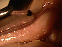
Expression, on the other hand, requires little cost, as it can be achieved with tools such as a Mastrota paddle (OcuSoft), double paddles (Bruder Healthcare), and the Meibomian Gland Evaluator™ (Johnson & Johnson Vision). Many years ago, I started with the Mastrota paddle, so for me that device has become routine in my clinic. Expressing the glands takes about 10 seconds—I take a paddle, place it behind the lower eyelid, and with my other thumb, push on the external lower eyelid with gentle pressure as I “milk” upward. Expression is graded as follows: Clear and thin like olive oil is normal, or grade 3; thickened or turbid is grade 2; paste-like is grade 1; and no expression is grade 0. Make sure to express the nasal to central lower eyelid so you get a good sampling of glands—aim for around 10. No anesthetic is necessary because if you have to press that hard to make a diagnosis, you already know the patient has MGD. Physiologically, the gentle pressure of a blink should express meibomian glands.
MGD is common and a primary reason for contact lens dropout.2 It is frequently found in glaucoma patients, especially those on prostaglandin analogs. It also presents in more than half of all patients undergoing cataract surgery.3 One study on children between the ages of four and 17 found that 42% of the young patients had meibomian gland atrophy.4 So rarely is there a time when this 10-second testing isn’t valuable.
Furthermore, treatment for evaporative dry eye, diagnosed via expression, is different from that used in aqueous deficient dry eye. You can’t provide effective treatment if you don’t know the type of dry eye, and you can’t determine the type of dry eye if you don’t express the meibomian glands. Make expression part of your routine eye exam, and you’ll quickly have a greater understanding of MGD and DED to more successfully manage your patient.
|
|
|
|
KEY TAKEAWAY: Research shows that 86% of all dry eye involves the lid margin and, specifically, meibomian gland dysfunction,1 so optometrists must express patients’ meibomian glands.
1. Lemp MA, Crews LA, Bron AJ, et al. Distribution of aqueous-deficient and evaporative dry eye in a clinic-based patient cohort: a retrospective study. Cornea. 2012 May;31(5):472-8.
2. Machalińska A, Zakrzewska A, Adamek B, et al. Comparison of morphological and functional meibomian gland characteristics between daily contact lens wearers and nonwearers. Cornea. 2015 Sep;34(9):1098-104.
3. Cochener B, Cassan A, Omiel L. Prevalence of meibomian gland dysfunction at time of cataract surgery. J Cata-ract Refract Surg. 2017 Feb;44(2):144-8.
4. Gupta P, Stevens MN, Priestley Y. Prevalence of meibomian gland atrophy in a pediatric population. Cornea. 2018 Apr;37(4):426-30.
|
|
|
|
| Supported by an independent medical grant from Kala Pharmaceuticals |
|
| |
| |
Review of Optometry® is published by the Review Group, a Division of Jobson Medical Information LLC (JMI), 19 Campus Boulevard, Newtown Square, PA 19073.
To subscribe to other JMI newsletters or to manage your subscription, click here.
To change your email address, reply to this email. Write "change of address" in the subject line. Make sure to provide us with your old and new address.
To ensure delivery, please be sure to add revoptom@lists.jobsonmail.com to your address book or safe senders list.
Click here if you do not want to receive future emails from Review of Optometry. |
|
|
|
|
|
|
|
|