| |
|
| Vol. 1, #05 • Thursday, October 15, 2020 |
|
|
|
|
| |
|
Review's Chief Clinical Editor
Paul M. Karpecki, OD, FAAO
Provides you with cutting-edge clinical strategies for optimal management of ocular surface disease and beyond.
|
|
|
|
|
|
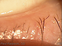 |
| A frothy tear film indicating saponified oils in a patient with early MGD. |
|
Research has shown that 86% of dry eye disease
(DED) has an evaporative or meibomian gland dysfunction (MGD) component.1
As such, one of the most important DED
pearls in this entire series is to be a great observer
of the eyelids.
Eye care providers must:
• Look for collarettes, the clear sleeves at the base of the lashes, as this is an indication of Demodex blepharitis and will lead to lash loss, inflammation and MGD.
• Scan for debris and discharge in the lashes, and especially biofilm along the lid margins indicating bacterial/Staphylococcal blepharitis.
• Express the meibomian glands of the lower nasal to central eyelid to ensure that clear oil is easily expressed. You can use a meibomian gland evaluator or a Mastrota paddle to assist. Abnormal expression is turbid, gelatinous/thickened, or paste-like; no expression is also abnormal.
• Look for froth on the eyelid margins or in the tear film, which would indicate saponified oils and a diagnosis of MGD.
• Watch the patient’s blink at the slit lamp to see if there is partial or incomplete closure.
• Determine if the patient’s eyelids don’t close properly at night by performing the K-B test. This involves having the patient close their eyes as if sleeping (not squeezing) and shining a penlight at the top of the closed eyelid to see if light escapes inferiorly.
• Finally, check for lid laxity, entropion, or ectropion.
Know that inflammation is often present when hyperosmolarity occurs, and almost all of the findings above will eventually lead to MGD, hyperosmolarity, and inflammation.
Thanks to colleagues like Donald Korb and Kelly Nichols, who have been decades ahead in the field of dry eye and meibomian gland dysfunction, we have come to understand the importance of the eyelids in dry eye disease.
In the next few clinical pearls, we’ll delve into a number of the above-mentioned eyelid contributors.
|
|
|
|
KEY TAKEAWAY: Research has shown that 86% of dry eye disease has an evaporative or MGD component so eye care providers must be a great observer of the eyelids.
1. Lemp MA, Crews LA, Bron AJ et al. Distribution of aqueous-deficient and evaporative dry eye in a clinic-based patient cohort: a retrospective study. Cornea. 2012 May;31(5):472-8
|
|
|
|
| Supported by an independent medical grant from Kala Pharmaceuticals |
|
| |
| |
Review of Optometry® is published by the Review Group, a Division of Jobson Medical Information LLC (JMI), 19 Campus Boulevard, Newtown Square, PA 19073.
To subscribe to other JMI newsletters or to manage your subscription, click here.
To change your email address, reply to this email. Write "change of address" in the subject line. Make sure to provide us with your old and new address.
To ensure delivery, please be sure to add revoptom@lists.jobsonmail.com to your address book or safe senders list.
Click here if you do not want to receive future emails from Review of Optometry. |
|
|
|
|
|
|
|
|