Submissions were graded in seven categories, each assigned a numerical value between 1 and 10: focus, exposure, field of view, difficulty of capturing image, absence of distracting elements, lighting and visual impact (wow! factor). The total scores from eight optometrist judges were averaged to determine the winners, presented below.
Special thanks to our judges: Amy Huddleston, Amanda Jimenez, Albert Nemiroff, Glenn Saxon, Mollie Saxon, Brooke Vegas, Karen Wadhams and Bryan Wolynski.
Grand Prize: Posterior Segment
Jessica Cameron, OD, FAAOUniversity of Florida Department of Ophthalmology, Gainesville, Florida
Special Acknowledgment: S. Gibran Khurshid MD, FACS , Retina and Ocular Oncology
Title: Solitary Retinal Capillary Hemangioma
Image System: Kowa Nonmyd Wx3D fundus camera
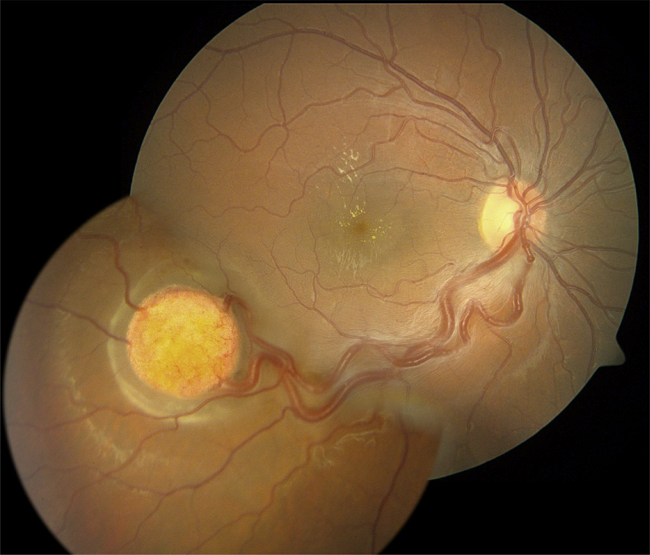 |
| Right eye with inferior temporal solitary retinal capillary hemangioma with feeder vessels and secondary macular exudation. Click image to enlarge. |
Grand Prize: Anterior Segment
Trey Sullins, OD
Sullins Eye Care, Starr Regional Medical Center, Athens, TN
Title: Emulsified Silicone Oil Droplets
Image System: Nikon FS3
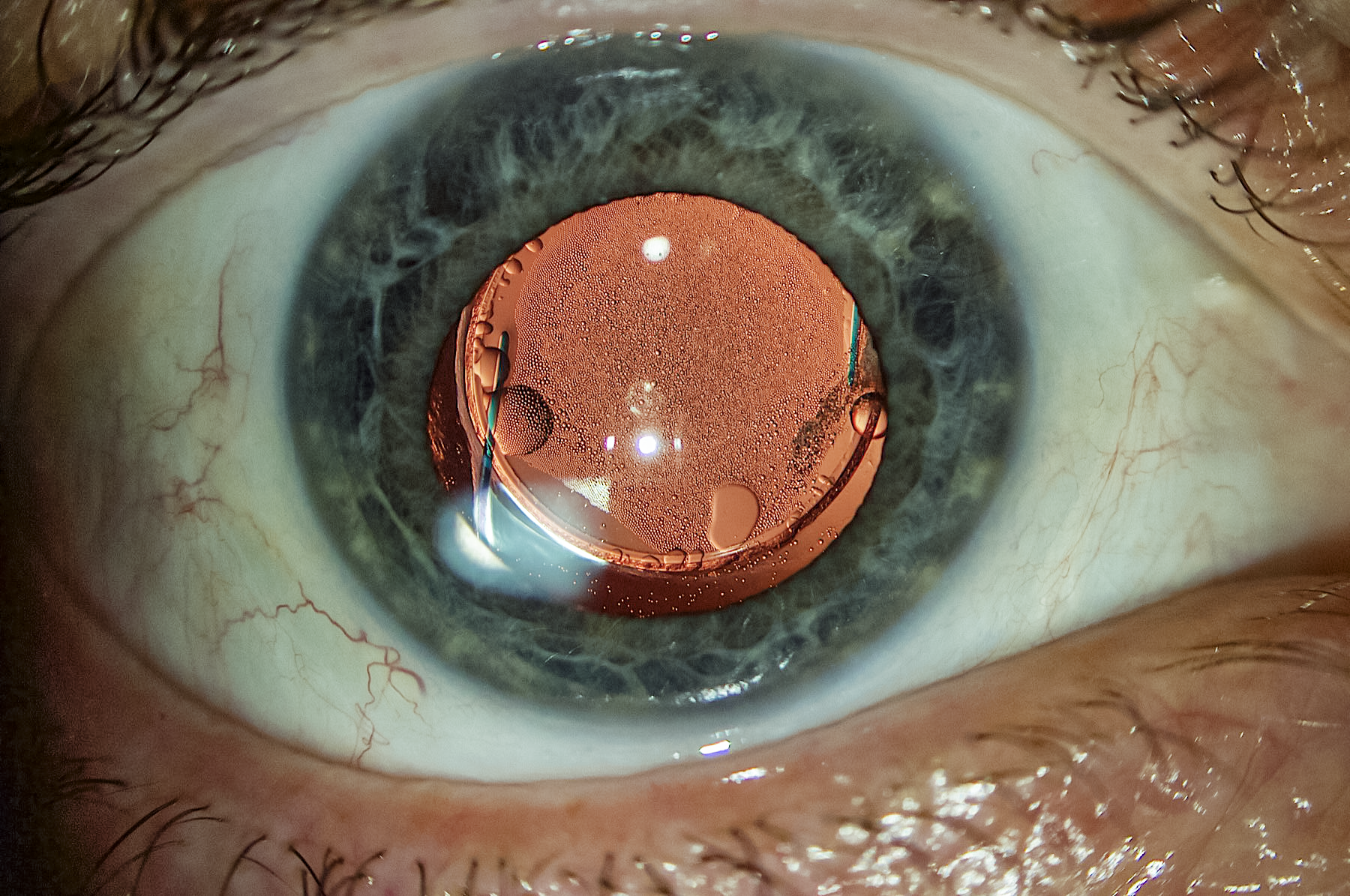 |
| Pseudophakic patient with giant retinal tear requiring silicone oil tamponade. The oil that was adherent to the PCIOL after removal from the vitreous cavity was easily aspirated from the IOL and capsular bag. Click image to enlarge. |
Honorable Mention 1
Kelley Sedlock, OD
Bennett and Bloom Eye Centers, Louisville, KY
Special Acknowledgment: Steve Bloom, MD
Title: Valsalva Retinopathy
Image System: Zeiss Visucam Pro
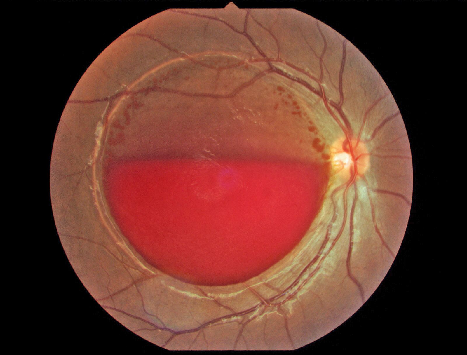 |
| The patient presented with sudden vision loss following delivery of her baby. There is a round detachment of the ILM throughout the macula with layered blood, which resolved over a period of 20 weeks. Click image to enlarge. |
Honorable Mention 2
Laura Lai Ming Chan, ODNortheastern State University Oklahoma College of Optometry, Tahlequah, OK
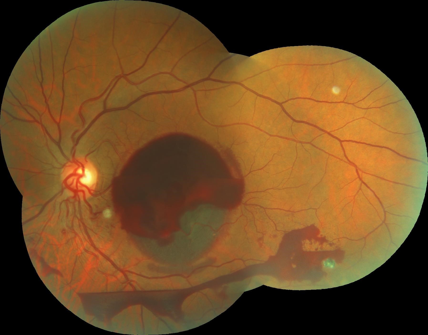
Title: Rhegmatogenous Retinal Detachment Associated with Macular Hole
Image System: Zeiss Clarus 500 Fundus Camera
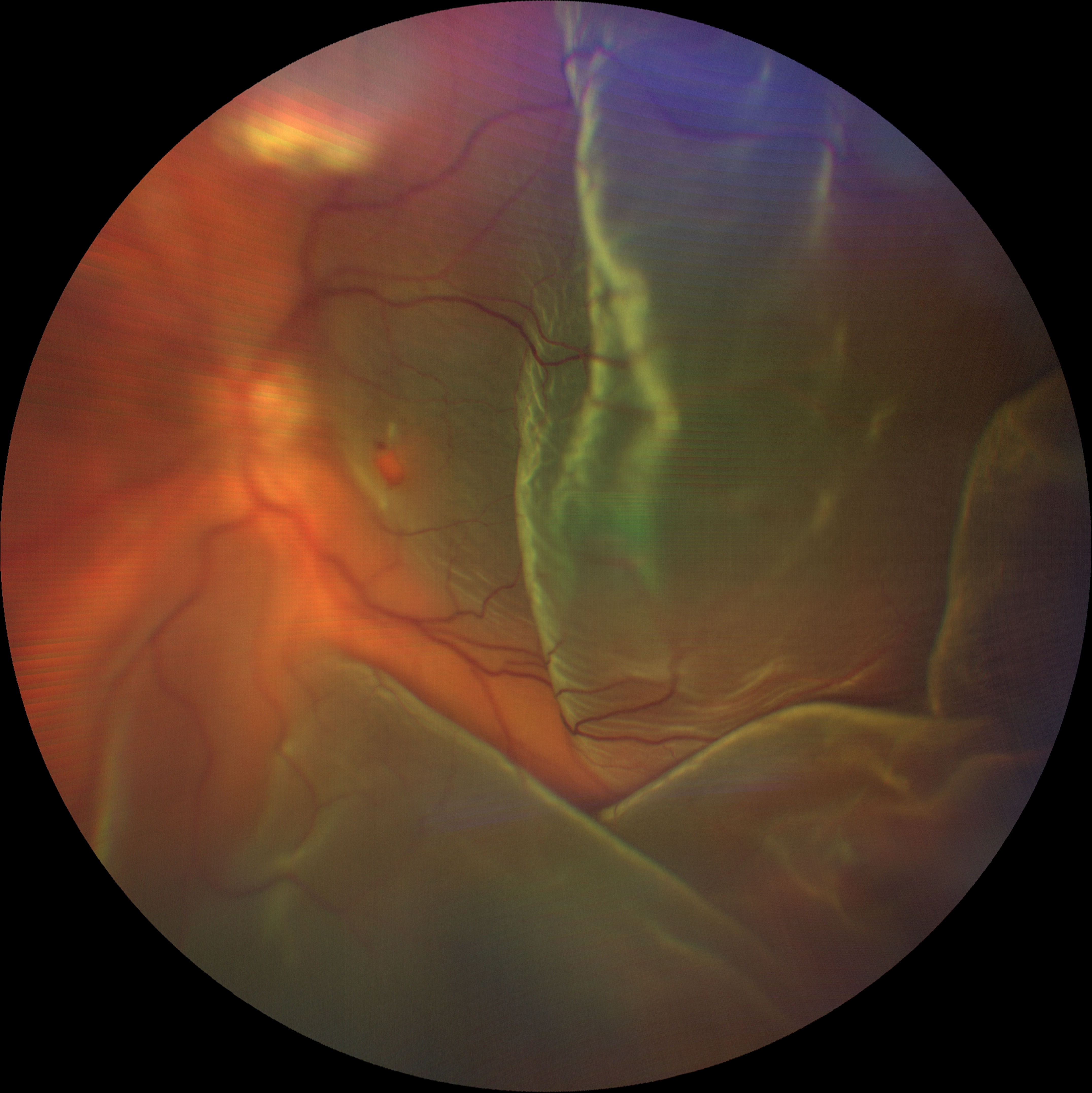 |
| A 61-year-old Native American female was diagnosed with rhegmatogenous retinal detachment associated with macular hole in her left eye. She was referred to a retina specialist for retinal detachment repair with pars plana vitrectomy. Recurrent retinal detachment with proliferative vitreoretinopathy was noted at her follow-up visit. Click image to enlarge. |
Honorable Mention 3
Corinne Casey, OD, FAAOKatzen Eye Group, Baltimore, MD
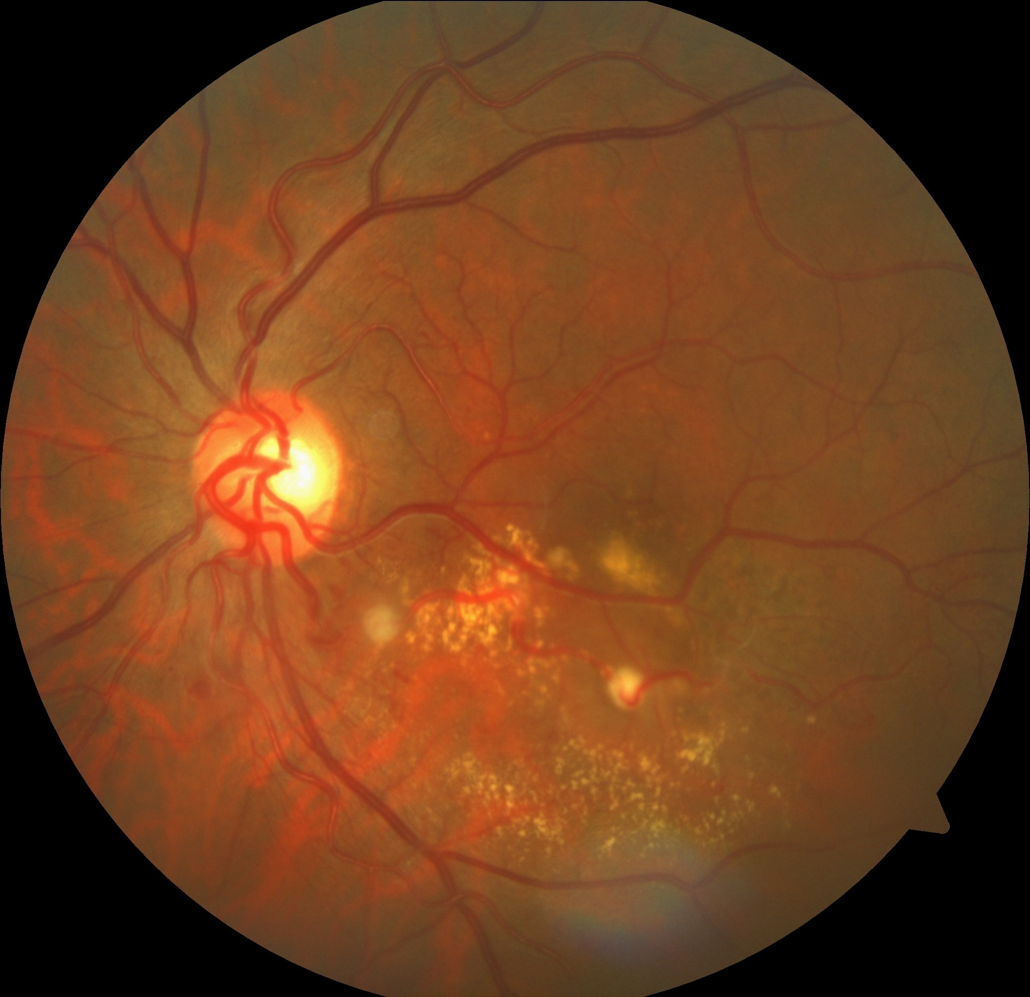
Title: Retinal Arterial Macroaneurysms
Image System: Zeiss Cirrus Photo 600
 |
 |
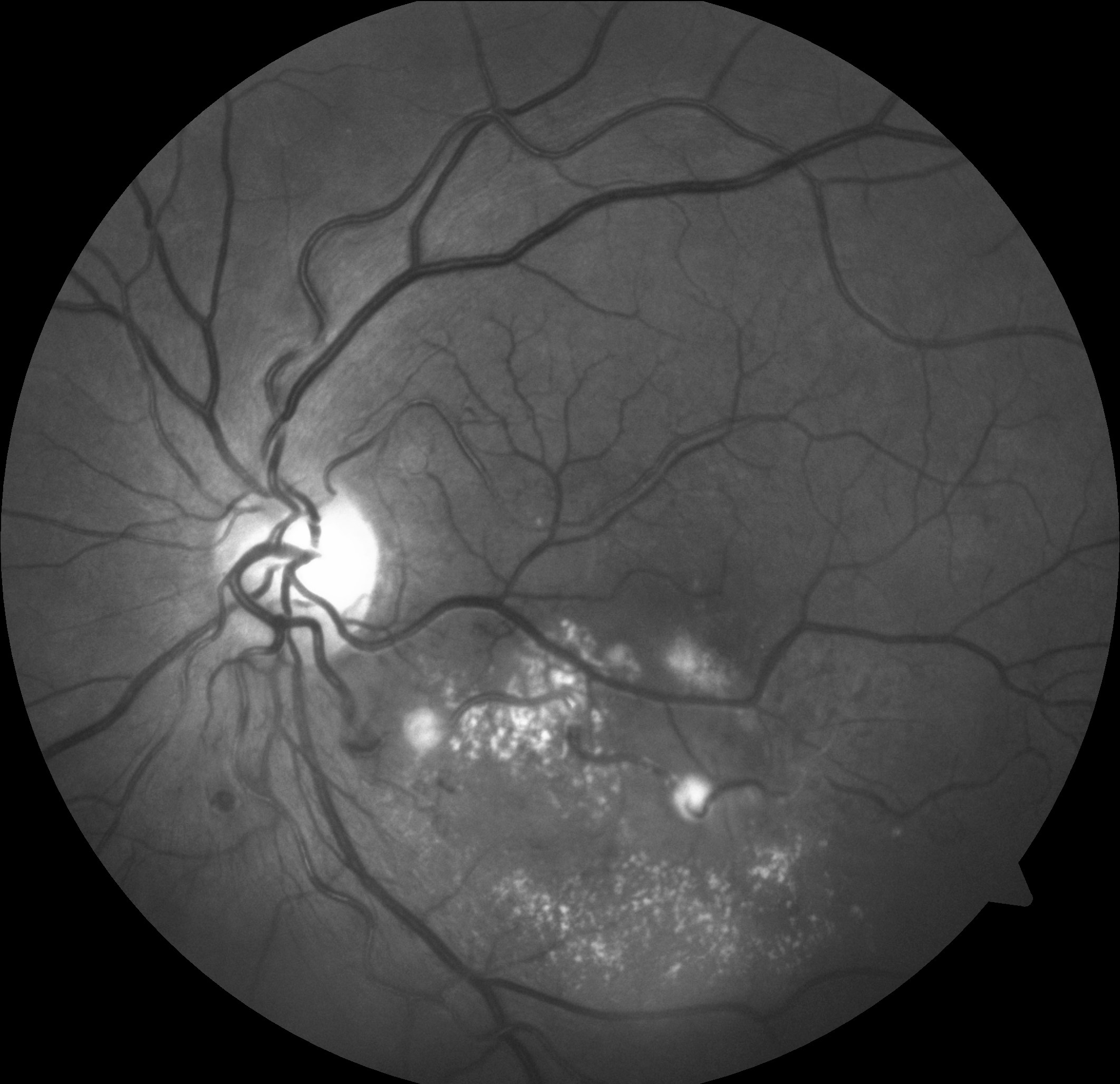 |
| A 63-year-old male presented with acute-onset central vision loss in the left eye secondary to a ruptured retinal arterial macroaneurysm. The characteristic layers of subretinal, intraretinal and preretinal hemorrhages resorbed within three months to reveal underlying exudates, sclerotic vessels and the fibrosed aneurysm. Two additional macroaneurysms, one of which is also fibrosed, are visible in the proximal portion of the inferotemporal arcade. Click images to enlarge. |
Honorable Mention 4
Corinne Casey, OD, FAAOKatzen Eye Group, Baltimore, MD
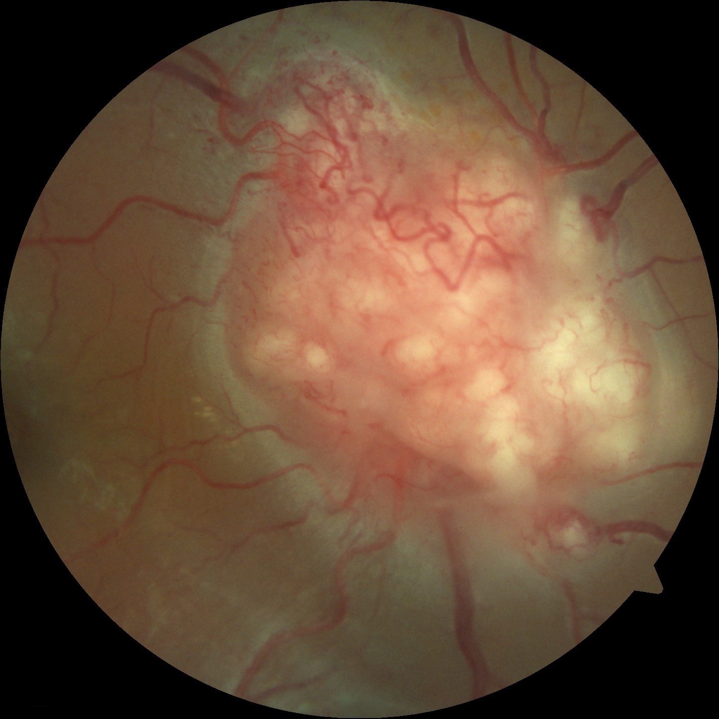
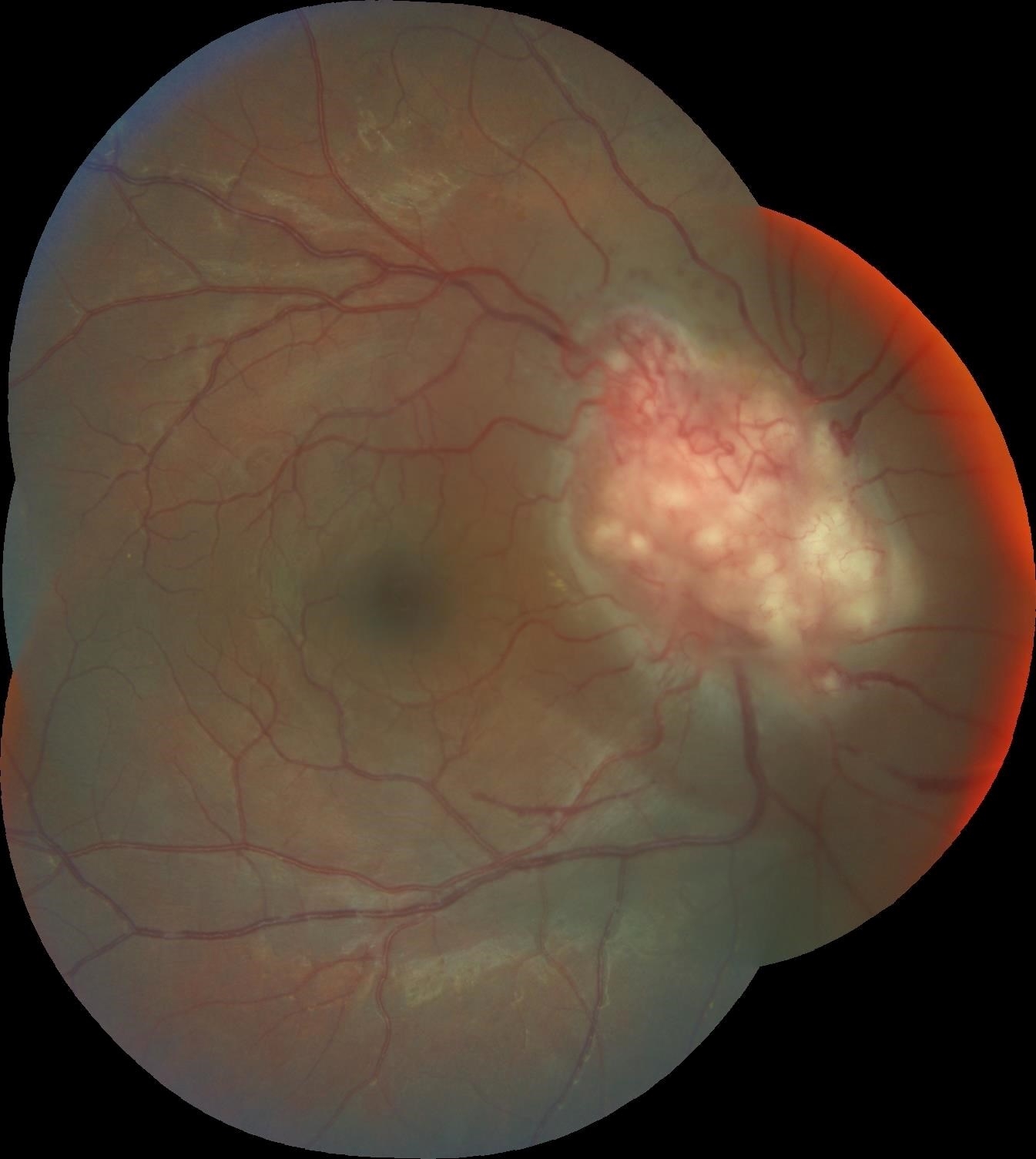
Title: Sarcoidosis-associated Optic Nerve Granuloma
Image System: Zeiss Cirrus Photo 600; Accutome B-scan Plus
 |
 |
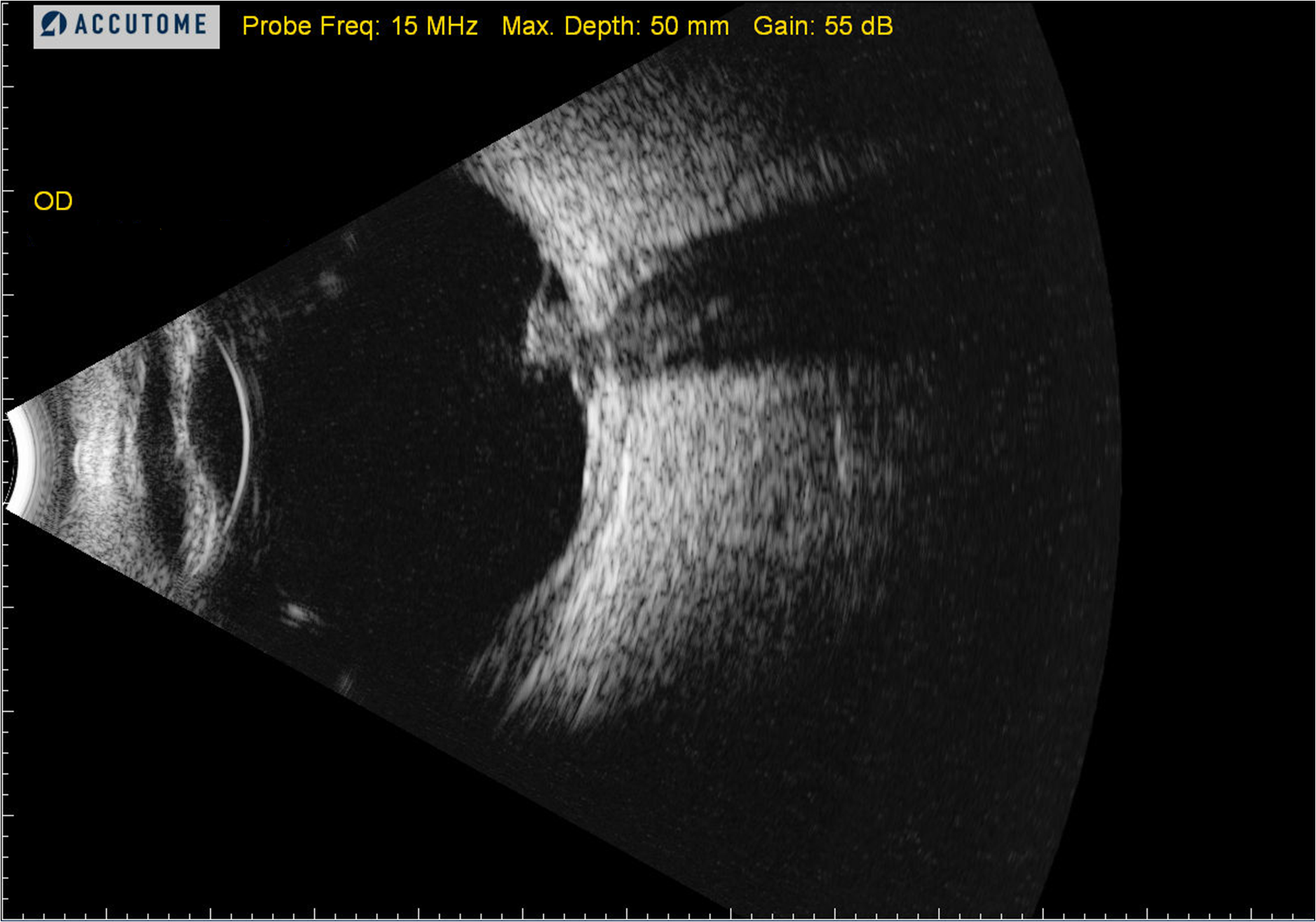 |
| A 25-year-old African American male presented with signs and symptoms of panuveitis in the left eye. His right eye was asymptomatic, yet dilated evaluation revealed an infiltrative optic nerve head mass. Further systemic workup, including chest x-ray and MRI of the brain and orbits, revealed widespread nodular opacities throughout the lungs and brain consistent with sarcoidosis. Click images to enlarge. |


