 A 56-year old white female transferred her care to my office in May 2005. She presented with an existing diagnosis of open-angle glaucoma, which was made 14 years earlier.
Her current ophthalmic medications included 1gtt Lumigan (bimatoprost, Allergan) O.U. h.s. and 1gtt 0.5% levobunolol O.U. b.i.d. Her systemic medications included estradiol and multivitmins. She reported allergies to Duracef (cefadroxil, Bristol-Myers Squibb) and penicillin.
Diagnostic Data
Upon initial presentation, she appeared adequately stabilized with Lumigan and levobunolol. Her IOP measured 11mmHg O.D. and 12mmHg O.S. Her best-corrected visual acuity was 20/20 O.D., O.S. and O.U. through hyperopic astigmatic, presbyopic correction. Pachymetry readings measured 497µm O.D. and 499µm O.S. Threshold visual fields demonstrated a stable, superior arcuate scotoma involving fixation O.D., and areas of superior field depression in the arcuate region not involving fixation O.S.
Her optic nerves were characterized by 0.85 x 0.90 cupping O.D. and 0.70 x 0.85 cupping O.S. Both nerves had severe thinning of the temporal neuroretinal rims (inferior greater than superior), with loss of all neuroretinal rim tissue O.D. from 6 o’clock to 7 o’clock. Her optic nerve characteristics were consistent with her visual field results. Gonioscopy of the anterior chamber angles demonstrated grade 2 to 3 open angles O.U., with minimal trabecular pigmentation.
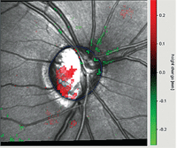
Topographic Change Analysis (on HRT-3) of our patient’s right eye demonstrated progressive nerve loss, as highlighted by the areas of red.
Her pupils were equally round and reactive to light and accommodation (ERRLA), with no afferent pupillary defect. Extraocular motilities were full in all positions of gaze.
Her anterior segments were unremarkable. Her crystalline lenses were clear O.U., and her internal examination was unremarkable except for glaucomatous optic neuropathy. The vitreous bodies were characterized by mild syneresis.
Then, in January 2009, the patient presented on an urgent basis with complaints of a tender, red left eye that had persisted for four days. On slit lamp examination, her right eye was uninvolved. The left eye, however, was characterized by a nasal area of sectoral episcleritis and an adjacent subconjunctival hemorrhage.
Discussion
Sectoral episcleritis is a common ocular inflammation of the episclera. It may be classified as diffuse, sectoral or nodular. Most cases readily respond to topical steroids. However, it is well documented that topical steroids have the propensity to increase intraocular pressure, especially after prolonged use. Furthermore, individuals with glaucoma, or those who are predisposed to glaucoma development, are more likely to experience elevated IOP secondary to topical steroid use. In these cases, steroid discontinuation will ultimately result in lowering IOP to baseline levels.
While many topical steroids are commercially available, most are designed to penetrate into the anterior chamber and quell ocular inflammation both outside the eye (e.g., episcleritis) and inside the eye (e.g., anterior uveitis). However, steroids that deeply penetrate the anterior chamber, such as dexamethasone and Pred Forte (prednisolone acetate, Allergan), are more likely to elevate IOP.
So-called “soft steroids,” such as Alrex (loteprednol etabonate 0.2%, Bausch & Lomb) and Lotemax (loteprednol etabonate 0.5%, Bausch & Lomb), are intended to reduce the likelihood of steroid responses. But, in some cases of aggressive ocular inflammation, these agents may not be as efficacious as other steroids.
One topical steroid that is not only effective against external inflammation, but also demonstrates reduced anterior chamber penetrence is fluorometholone. Fluorometholone is very useful in glaucoma patients with external ocular inflammations, such as episcleritis, and it demonstrates a low incidence of elevated IOP.
So, in order to treat this patient’s episcleritis, I prescribed FML Forte (fluorometholone 0.25%, Allergan) q2h (while awake) for two days, then q.i.d. for four days. When the patient returned in four days, as requested, she reported significant relief O.S. The episclera was markedly improved, and her IOP measured 12mm Hg O.D. and 13mm Hg O.S. We tapered the steroid during the next few days until complete resolution.
End of story, right? Not quite.
On November 20, she presented to my partner with acute complaints of bilateral redness, discomfort and blurred vision. When questioned, she reported that her discomfort had elevated in the last “week or so.” She was still taking Lumigan and levobunolol, and said that she had used the FML Forte “occasionally.” This raised a red flag, because I always inform patients that they should not use topical steroid drops indiscriminately due to the risk of potential side effects.
At this visit, both lids were moderately swollen, and there was moderate diffuse episcleral injection O.U. Both anterior chambers were quiet. Both corneas were slightly hazy, which resulted in acuity reduction to 20/30 O.U. Pupils were ERRLA, with no afferent pupillary defect. IOP measured 30mm Hg O.U.
My partner was concerned about medicamentosa and a steroid response. So, he discontinued the Lumigan, levobunolol and FML Forte, and started her on a regimen of Travatan Z (travoprost, Alcon) 1gtt O.U. h.s., Lotemax b.i.d. O.U., and preservative-free tears q.i.d. O.U. He also instilled two drops of Durezol (difluprednate, Sirion Therapeutics) in the office, and instructed her to return in three to four days for follow-up with me.
She returned on November 24 and reported good compliance with her new regimen, but complained of increasing pain and blurrier vision, as well as an episodic complaint of severe pain O.S.
At this visit, her IOP measured 35mm Hg O.D. and 39mm Hg O.S. The episclera was mildly injected bilaterally, and the anterior chambers remained deep and quiet. Heidelberg Retina Tomography-3 (HRT-3, Heidelberg Engineering) images clearly demonstrated progressive neuroretinal rim loss inferotemporally O.D. (O.S. remained stable).
So, how do we now manage this patient? On pointed questioning, the patient admitted using the FML Forte “off and on” for the past several months, and began using it once daily about two months prior. But, after experiencing little to no relief, she gradually increased the dosage to q.i.d. until her visit on November 20.
I told the patient in no uncertain terms that self-medicating with the steroid had resulted in a complex set of problems, including moderately elevated IOP in the presence of severely compromised optic nerves, resultant decreased vision, anterior segment inflammation and pain. I also expressed my concern that she may experience permanent vision loss in her right eye.
While there are many ways to approach a patient in this situation, it is important to realize that this situation will not simply quiet down and resolve in just a few days. On one hand, increasing and pulsing topical steroids may result in quieting the eye sooner, but it will likely cause further elevated IOP, which could compromise her fragile neuroretinal rims.
On the other hand, a passive approach may delay quieting of the inflammation, which could result in more anterior chamber and/or trabecular inflammation. Furthermore, it would be exceedingly difficult to lower IOP in the presence of significant anterior segment inflammation.
The patient has a well-documented history of tolerating Lumigan and levobunolol, so I instructed her to restart both medications and discontinue the Travatan-Z. I also asked her to continue the Lotemax b.i.d. I would be surprised if the Lotemax doesn’t elevate IOP; however, I am concerned about the potential for rebound inflammation secondary to withdrawal of the FML Forte. I decided not to add another topical ocular hypotensive agent, as it may perpetuate the inflammatory nature of her problem.
Our patient is scheduled to be seen again in two weeks––sooner, if she experiences any further decrease in vision. I explained that I did not expect her to have any significant improvement in her vision before the next visit, because her IOP will likely remain high.
With time, the inflammation will likely quiet and her IOP may decrease modestly. If her IOP remains elevated at follow-up, I will medicate her with oral Diamox (acetazolamide, Lederle Laboratories) and may recommend a quick tapering of the topical steroids.
