 A 67-year-old white male had recently undergone uncomplicated cataract extraction in his right eye and was presenting for a three-week postoperative visit. He had undergone cataract extraction in his fellow eye three years earlier and had experienced a somewhat protracted course of postoperative inflammation at that time.
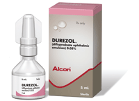
That prolonged postoperative inflammation occurred while the patient was using prednisolone acetate 1%. So this time, he was prescribed Durezol (difluprednate ophthalmic emulsion 0.05%, Alcon), which he has been using as directed.
At the one-day and one-week postoperative visits, the patient remarked that he felt great, with virtually no discomfort. At today’s visit, however, he said his vision seemed a bit blurry, and he was experiencing what he described as “a pressure sensation.”
Best-corrected visual acuity was 20/25 O.D. and 20/20 O.S. Biomicroscopic examination revealed diffuse mild corneal edema, few inflammatory cells and no flare in an otherwise white and quiet eye. At this visit, intraocular pressure was 39mm Hg O.D. and 16mm Hg O.S. At his previous postoperative visits, IOP was 14mm Hg and 16mm Hg respectively. Gonioscopy revealed completely open anterior chamber angles O.U. with no trabecular obstructions or other abnormalities. At this point, the patient was diagnosed with a steroid-induced IOP elevation.
This month, we’ll examine steroid-induced glaucoma, as well as a potent new topical steroid.

Steroid-Induced Glaucoma
While topical ophthalmic corticosteroids are th-ose that are most likely to precipitate a rise in IOP, other modalities of steroid use include intraocular and periocular injected steroids, topical periocular dermatological creams, inhaled steroids and oral steroids.1-13 In cases in which a rise in IOP occurs, the duration of steroid use tends to be weeks to months, frequently results from inappropriate use, and may even stem from improper clinical follow-up.
But, IOP elevation can occur after shorter periods of time even when steroids are used appropriately and under close observation by the clinician. Such steroids as dexamethasone or prednisolone are more likely to cause a rise in IOP than those such as loteprednol or fluorometholone due to their penetrance and glucocorticoid activity.3,14
Steroid-induced glaucoma presents with an open anterior chamber angle, and the rise in IOP appears to be due to outflow reduction. Beyond this, very little can be conclusively said regarding the condition’s pathophysiology. One theory blames steroids for the accumulation of glycosaminoglycans in the trabecular meshwork. Once hydrated, glycosaminoglycans cause an aqueous outflow obstruction.15 Another hypothesis holds that steroids decrease the phagocytic ability of the trabecular meshwork endothelial cells, resulting in an increase in debris and decrease in aqueous processing ability.3
Genetic mapping has recently led to greater understanding of steroid-induced glaucoma. Steroids are believed to induce the expression of a gene located on chromosome 1, known as TIGR or GLCIA. The resultant genetic product is a glycoprotein called myocilin.5 In the trabecular meshwork, myocilin is found in the cytoplasm of the cells and in the juxtacanalicular region in association with fibrillar extracellular matrix components. This increase in myocilin subsequently increases outflow resistance.16
Management of patients with steroid-induced glaucoma involves discontinuation of the precipitating medication, if possible. When steroid treatment is short-term, discontinuation usually results in a return to pre-treatment IOP levels.5 Patients undergoing steroid treatment over several continuous months to years, however, may develop a chronic IOP elevation that is unalterable by steroid cessation.17
In such cases, IOP elevation can be treated with topical aqueous suppressants. In that the dysfunction may be due to increased resistance to aqueous outflow at the trabecular meshwork, therapies designed to increase trabecular outflow, such as laser trabeculoplasty and miotics, are of questionable benefit. However, prostaglandin analogs (PGAs) appear to be successful in the management of steroid-induced glaucoma.18 However, PGAs may be contraindicated by the underlying inflammatory process that necessitated steroid use in the first place.
What is Durezol?
Durezol is a difluorinated derivative of prednisolone with potent anti-inflammatory activity. Difluprednate was engineered to be more potent than its progenitor through the addition of two fluorine atoms to the initial prednisolone molecule. Additional modifications increased corneal penetration, increased glucocorticoid receptor affinity and enhanced its anti-inflammatory activity.19,20
In 2008, the U.S. Food and Drug Administration approved Durezol for the treatment of postoperative inflammation and pain associated with ocular surgery.21 In fact, it was the first steroid approved specifically for pain management. The recommended dosing regimen is one drop, two to four times daily beginning 24 hours after surgery and continuing throughout the first two weeks of the postoperative period, followed by two times daily for a week with tapering thereafter based upon clinical response.
The lipid emulsion has a smaller particle size, which increases bioavailability, provids uniform medication concentration in each drop, and eliminates the need for shaking before use.20
Like any topical steroid, Durezol has the potential to increase IOP. But, its safety profile is quite good—only 3% of patients experienced IOP elevation, similar to that seen in other topical steroids.21 Durezol has been shown to be clinically comparable to the very potent betamethasone for postoperative inflammation with an equal likelihood of a steroid response.22 Due to its enhanced bioavailability and clinical potency, Durezol tends to need less frequent dosing than other topical steroids.
In the case of the patient presented here, once we made the diagnosis, we educated him on the condition and instituted a tapering regimen. Additionally, the patient was temporarily prescribed Combigan (timolol 0.5%/brimonidine 0.20%, Allergan) b.i.d. O.D. When the patient returned as scheduled two days later, his IOP was reduced to 14mm Hg O.D., and he felt fine.
While IOP elevation can occur from the use of topical steroids, such as Durezol, the clinician can easily manage the situation through appropriate follow-up and use of IOP-reducing medications as needed.
At this time, Durezol is indicated for the management of postoperative pain and inflammation in ocular surgery. However, studies have also demonstrated its efficacy and uses in other conditions, such as anterior uveitis.
Drs. Sowka and Kabat are consultants for Alcon. They have no direct financial interests in any products mentioned.
1. Mitchell P, Cumming RG, Mackey DA. Inhaled corticosteroids, family history, and risk of glaucoma. Ophthalmology. 1999 Dec;106(12):2301-6.
2. Mohan R, Muralidharan AR. Steroid induced glaucoma and cataract. Indian J Ophthalmol. 1989 Jan-Mar;37(1):13-6.
3. Kong L, Zhang C, Chen M, Xue G. Clinical analysis of steroid glaucoma. Yan Ke Xue Bao. 1995 Mar;11(1):53-6.
4. Baratz KH, Hattenhauer MG. Indiscriminate use of corticosteroid-containing eyedrops. Mayo Clin Proc. 1999 Apr;74(4):362-6.
5. Sapir-Pichhadze R, Blumenthal EZ. Steroid induced glaucoma. Harefuah. 2003 Feb;142(2):137-40.
6. Singh IP, Ahmad SI, Yeh D, et al. Early rapid rise in intraocular pressure after intravitreal triamcinolone acetonide injection. Am J Ophthalmol. 2004 Aug;138(2):286-7.
7. Detry-Morel M, Escarmelle A, Hermans I. Refractory ocular hypertension secondary to intravitreal injection of triamcinolone acetonide. Bull Soc Belge Ophtalmol. 2004;(292):45-51.
8. Kaushik S, Gupta V, Gupta A, et al. Intractable glaucoma following intravitreal triamcinolone in central retinal vein occlusion. Am J Ophthalmol. 2004 Apr;137(4):758-60.
9. Akduman L, Kolker AE, Black DL, et al. Treatment of persistent glaucoma secondary to periocular corticosteroids. Am J Ophthalmol. 1996 Aug;122(2):275-7.
10. Garrott HM, Walland MJ. Glaucoma from topical corticosteroids to the eyelids. Clin Experiment Ophthalmol. 2004 Apr;32(2):224-6.
11. Thoe Schwartzenberg GW, Buys YM. Glaucoma secondary to topical use of steroid cream. Can J Ophthalmol. 1999 Jun;34(4):222-5.
12. Desnoeck M, Casteels I, Casteels K. Intraocular pressure elevation in a child due to the use of inhalation steroids—a case report. Bull Soc Belge Ophtalmol. 2001;(280):97-100.
13. Caldwell JR, Furst DE. The efficacy and safety of low-dose corticosteroids for rheumatoid arthritis. Semin Arthritis Rheum. 1991 Aug;21(1):1-11.
14. Ilyas H, Slonim CB, Braswell GR, et al. Long-term safety of loteprednol etabonate 0.2% in the treatment of seasonal and perennial allergic conjunctivitis. Eye Contact Lens. 2004 Jan;30(1):10-3.
15. Sherwood M, Richardson TM. Evidence for in vivo phagocytosis by trabecular endothelial cells. Invest Ophthalmol. 1958; 59:216.
16. Ohlmann A, Tamm ER. The role of myocilin in the pathogenesis of primary open-angle glaucoma. Ophthalmologe. 2002 Sep;99(9):672-7.
17. Espildora J, Vicuna P, Diaz E. Cortisone induced glaucoma: A report on 44 affected eyes. J F Optalmol. 1981;4:503-8.
18. Scherer WJ, Hauber FA. Effect of latanoprost on intraocular pressure in steroid-induced glaucoma. J Glaucoma. 2000 Apr;9(2):179-82.
19. Bikowski J, Pillai R, Shroot B. The position not the presence of the halogen in corticosteroids influences potency and side effects. J Drugs Dermatol. 2006 Feb;5(2):125-30.
20. Yamaguchi M, Yasueda S, Isowaki A, et al. Formulation of an ophthalmic lipid emulsion containing an anti-inflammatory steroidal drug, difluprednate. Int J Pharm. 2005 Sep;301(1-2):121-8.
21. Korenfeld M, Silverstein S, Cook D, et al. Difluprednate ophthalmic emulsion 0.05% for postoperative inflammation and pain. J Cataract Refract Surg. 2009 Jan;35(1):26-34.
22. Jamal KN, Callanan DG. The role of difluprednate ophthalmic emulsion in clinical practice. Clin Ophthalmol. 2009;3:381-90.

