 Q: Even though the worst of the Acanthamoeba epidemic may be considered behind us, how can I care for my patients and lessen their risk of exposure or infection?
A: The practitioner needs to assess each patients willingness and ability to comply with wear regimens, says Elmer Tu, M.D., director of the Cornea and External Disease Service at the
The lens care regimen that you recommend to the patient will play a role in his or her chances of exposure. In a recent study, hydrogen peroxide systems showed the highest kill rates over six hours vs.
multipurpose solutions.2,3 But in general, Acanthamoeba cysts and trophozoites dont adhere tenaciously to lens surfaces and may be removed with a vigorous rub and rinse.
Maintaining excellent hygiene and keeping lenses as clean as possible are essential practices, says Charlotte Joslin, O.D., of the

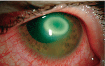
This Acanthamoeba patient presented with a late-forming ring infiltrate and contiguous scleritis. Photo courtesy: Christine W. Sindt, O.D.
Dont forget to discuss case hygiene with the patient. In a contaminated lens case, cysts can survive in the biofilm. A case can become contaminated in as little as one week, with contamination rates as high as 60%.4 The tendency of the most common strain of Acanthamoeba is to aggregate upon encountering a cleaning solution, and it is this tendency that may make it harder to eradicate amoeba from the lens case.4 When cleaning the case, specify that the patient cleans the upper inner rim of the case, where contamination is most likely.4
When a patient experiences any sign of infection, such as persistent pain, decreased vision, redness or photophobia, make sure that he or she contacts you and comes in for an examination right away.
When it comes to diagnosis, a confocal microscope may provide the best results, especially if the practitioner has experience in identifying Acanthamoeba.2,5 Culturing is another method that will bring definite results; however, this is often time consuming.2,5 Using both methods in tandem may bring the highest rates of diagnostic success, though the confocal microscope is definite enough to allow treatment to begin.2,5
Patient Consultation Checklist
Each of these factors, when properly conveyed to and understood by the patient, can play a role in his or her ability to lessen exposure to Acanthamoeba. So, make sure to include each of these topics in your contact lens consultationdont assume that the patient already knows this information!6
Create personalized case replacement schedules.
Provide a compliance checklist. Consider including a signed agreement between you and the patient.
Make sure patients (especially those who wear extended-duration lenses) understand the importance of regular replacement.
Educate patients about the clinical signs and symptoms of possible infection.
Remind patients regularly that contact lenses are a prescription medical device.
Ensure that the patients lenses and solutions are compatible.
1. Shovlin JP, ed. Accounting for Acanthamoeba. Rev Optom 2007 Sep;144(9):133.
2. Cavanagh HD, Shovlin JP, Sindt CW. All about Acanthamoeba. Rev Cornea Contact Lens 2009 Jan/Feb; 146(1):15.
3. Shoff ME, Joslin CE, Tu EY, et al. Efficacy of contact lens systems against recent clinical and tap water Acanthamoeba isolates. Cornea 2008 Jul;27(6):713-9.
4. Shovlin JP. Report from ARVO: Cornea. Rev Optom 2009 May;146(5):43-8.
5. Tu EY, Joslin CE, Sugar J, et al. The relative value of confocal microscopy and superficial corneal scrapings in the diagnosis of Acanthamoeba keratitis. Cornea 2008 Aug; 27(7):764-72.
6. Shovlin JP, ed. Protect patients against Acanthamoeba. Rev Optom 2007 Oct;144(10):113.

