A 53-year-old Haitian female presented complaining that she “couldn’t see well” and that her eyes were “not good.” She reported a six months prior visit for a mosquito bite on her eyelid that required lancing. Otherwise, her ocular history was unremarkable. She had never worn glasses.

Her medical history was significant for HIV (she was diagnosed about seven months earlier). She was taking HIV medications, but did not know their names. She also did not know her CD4 count or viral load.
On examination, her uncorrected visual acuity measured 20/20 O.U. at distance and near (she read J5 at near O.U.). Confrontation fields were full to careful finger counting O.U. The pupils were equally round and reactive, with no afferent pupillary defect. The anterior segment was unremarkable in both eyes. The anterior vitreous was clear. Her intraocular pressure measured 15mm Hg O.U.
Dilated fundus exam showed a clear vitreous O.U. The optic nerves appeared healthy, and she had small cups with good rim coloration and perfusion. We noted a small flame-shaped hemorrhage near her right macula. The peripheral retina of each eye exhibited notable changes (Figures 1 and 2). We also obtained an OCT scan of her maculae (Figures 3 and 4).
Take the Retina Quiz
1. What do the peripheral retinal changes represent?
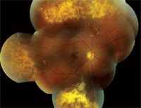
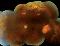
1, 2. Here are posterior pole and peripheral fundus montages of the right and left eyes of our patient (O.D. left, O.S. right). Note the peripheral retinal changes. Of particular interest are subtle changes seen superiorly O.D. and temporally O.S. What do these represent?
a. Chorioretinal scars.
b. Active chorioretinitis.
c. Active retinitis.
d. Inactive retinitis.
2. What is the likely diagnosis?
a. Toxoplasmosis.
b. Acute retinal necrosis (ARN).
c. Burned out cytomegalovirus (CMV) retinitis.
d. Progressive outer retinal necrosis (PORN).
3. What do the two peculiar changes seen superior O.D. and temporally O.S. likely represent?
a. Hemorrhagic retinal pigment epithelium (RPE) detachment.
b. Exudative retinal detachment.
c. Eccentric disciform process.
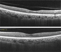
3, 4. What do her OCT scans reveal (O.D. top, O.S. bottom)?
d. Choroidal rupture.
4. What does the OCT of the macula show?
a. Nothing clinically remarkable.
b. Serous detachment.
c. RPE detachment.
d. Choroidal neovascularization (CNV).
5. How should this patient be managed?
a. Observation.
b. Referral to infectious disease specialist.
c. Begin anti-viral therapy.
d. Begin antibiotic therapy.
For answers, see below.
Discussion
The peripheral retinal changes seen in our patient likely represent burned out cytomegalovirus retinitis. It is not active at this time. In our patient, it is seen as widespread areas of circumferential chorioretinal scarring in the absence of vitreous cells. Active CMV would appear whiter with primary involvement in the superficial retina. But, in our patient, the scarring involves all the layers of the retina including the RPE and choroid.
CMV retinitis may be present anywhere in the retina. Posterior pole lesions have a characteristic white, hemorrhagic appearance with retinal necrosis and edema, while the peripheral lesions are more indolent and nonhemorrha-gic. Additionally, vitreous and anterior chamber cells will be present. The anterior chamber inflammation may represent spillover from the vitreous. As the CMV begins to resolve, the borders of the lesions become granular and grayer in color.
CMV retinitis is the most common ocular complication that is seen in AIDS patients. CMV retinitis develops in patients whose CD4+ T-cell counts are slightly to significantly below 50. In the early days of the AIDS epidemic, CMV was very common, developing in up to 40% of patients. But, with the advent of HAART (highly active antiretroviral therapy), CMV has virtually disappeared––with a 55% to 95% decline in the number of CMV retinitis cases.1
HAART therapy consists of a combination of nucleoside reverse transcriptase inhibitors, non-nucleoside reverse transcriptase inhibitors and at least one protease inhibitor. It is now considered the standard treatment for HIV.
Our patient did not know her CD4 counts or her viral load, but we can assume that it was low when she was diagnosed seven months ago. Undoubtedly, she was placed on HAART therapy and her immune system was able to overcome the active infection.
So, is there a possibility that this represents something else? Without definitive diagnostic testing, it is impossible to know for sure that this represents old CMV. Because of its peripheral location and circumferential pattern, it is possible that this could also represent old acute retinal necrosis (ARN), a viral infection that is caused by the herpes simplex family. ARN tends to be rapidly progressive and non-self-limiting, often resulting in retinal detachment.
The peripharal exudative lesions are of particular interest. The lesion in the right eye is subtler and can be seen just posterior to the area of chorioretinal scarring. There appears to be a shallow hemorrhagic detachment of the RPE and associated circinate exudate. The lesion in the temporal retinal of the left eye is larger and more elevated. It also represents a hemorrhagic detachment of the RPE, but the blood is beginning to become dehemoglobinized—evidenced by the white appearance in the center.
These lesions are most peculiar and atypical of what is customarily seen in CMV or ARN. Could it represent another disease process entirely? Possible causes include polypoidal choroidal vasculopathy (PCV), a condition that results in aneurismal dilations of the choroid vessels resulting in episodic bleeding and scarring, or eccentric disciform process, a CNV that is located outside the posterior pole and macula. Eccentric disciforms are rare, isolated and usually do not include multiple lesions, whereas PCV tends to involve more of the posterior pole.
Bottom line, we are uncertain about the exact etiology of the lesions. Regardless, because of their peripheral location, the lesions do not appear to be a threat to her macula or visual function. As a result, we recommended close observation and instructed her to return in four to six weeks. We also requested a report from her physician with regards to her current medications and immune status.
Answers to the Retina Quiz
1. d
2. c
3. a
4. a
5. a
1. Jabs DA, Van Natta ML, Kempen JH, et al. Characteristics of patients with cytomegalovirus retinitis in the era of highly active antiretroviral therapy. Am J Ophthalmol. 2002 Jan;133(1):48-61.
