 A 48-year-old black male presented with a three-month history of reduced visual acuity and discomfort in his right eye. He reported that, over the past month, his vision had declined to the point that he could “hardly see anything.” The left eye seemed fine. He never wore glasses.
His medical history was significant for HIV. He reported taking medications for the condition, but was uncertain of their names.
His entering visual acuity was hand motion O.D. and 20/200 O.S. Upon hyperopic correction, his acuity improved to 20/20 O.S. Confrontation fields in the left eye were full to careful finger counting. His pupils were equally round and reactive, with a 1+ afferent defect O.D. IOP measured 17mm Hg O.D. and 12mm Hg O.S.
The anterior segment evaluation of the right eye revealed significant changes (figure 1). The anterior segment examination of the left eye was completely unremarkable.
The right fundus appeared extremely hazy; however, changes could be seen in the posterior pole and superior nasal area (figure 2). The fundus examination of the left eye was completely normal.
Take the Retina Quiz

1. Anterior segment image of our patient’s right eye. What do you notice?
1. What do the changes seen on the right cornea represent?
a. Gutta.
b. Keratic precipitates (KP).
c. Infectious infiltrates.
d. Ulcerative keratitis.
2. How would you classify the clinical presentation of the right eye?
a. Retinochoroiditis.
b. Endophthalmitis.
c. Posterior uveitis with anterior segment spillover.
d. Panuveitis.
3. Which screening test could yield a positive result in our patient?
a. Fluorescent treponemal antibody-absorption (FTA-ABS).
b. Rapid plasma reagin (RPR).
c. Toxoplasmosis IgM and IgG.
d. All of the above.
4. Based upon the patient history and clinical findings, what is the most likely diagnosis?
a. Active cytomegalovirus (CMV).
b. Active toxoplasmosis.
c. Active ocular syphilis.
d. All of the above.
5. How should this patient be managed initially?
a. Topical steroids and cycloplegics.
b. Topical steroids.
c. NSAIDs.
d. Intravitreal antibiotic injection.
For answers, see below
DiscussionOur patient had a panuveitis, which represents an inflammation of the entire uveal tract. Indeed, he exhibited an active anterior uveitis with cells in the anterior chamber and obvious KP on the endothelium. There also were cells in the anterior and posterior vitreous, which made the view of the retina appear very hazy. Finally, there was an active chorioretinal lesion that involved the macula as well as a less-active lesion located superior nasal O.D.
In his history, he indicated that the vision in his left eye had been reduced for at least three months. That explains the macular lesion; however, the more peripheral lesion would not cause the central vision loss that he experienced at the time of his examination. Additionally, given the amount of pigment surrounding the lesion, we speculated that it had been present for longer than three months.
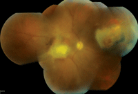
2. A wide-angle view of our patient’s right eye.
So, what is the underlying cause? At first glance, the retinal lesions looked to be characteristic of active toxoplasmosis. To confirm our suspicions, we ordered a blood work-up that included a CBC and serology to rule out syphilis (FTA-ABS and RPR); toxoplasmosis IgG and IgM antibodies; a chest X-ray; and rheumatoid factor, antinuclear antibody and an angiotensin converting enzyme (ACE) testing to rule out the presence of other autoimmune diseases.
CMV retinitis was not really a consideration. Even though we didn’t know his CD4 count, the amount of inflammation that we observed in our patient was not consistent with CMV. Traditionally, CMV occurs when the immune system is severely compromised. So, when patients develop CMV, their immune system is not strong enough to mount the kind of inflammatory response that we noted in our patient.
The results of the blood work didn’t make it any easier. Both blood tests for syphilis were positive. Additionally, one of the tests for toxoplasmosis was positive (the IgG), indicating that he had been exposed to toxoplasmosis. (Fortunately, the IgM test was negative, suggesting that the toxoplasmosis infection was not recent.) And, if that wasn’t confusing enough, the ACE test also came back as mildly positive.
So, could it have been that our patient had multiple active conditions? It was fairly unlikely. Without question, however, he did have active syphilis. Because syphilis is considered the “great masquerader,” it certainly could have been the cause of his panuveitis. But, it was also possible that toxoplasmosis was the root cause––especially given the positive serology and the characteristic clinical appearance.
Both syphilis and toxoplasmosis are caused by parasites, so it seems logical that associated lesions exhibit a similar clinical appearance.
• Toxoplasmosis is caused by an intracellular parasite, Toxoplasma gondii. Active toxoplasmosis retinochoroiditis typically presents as a feathery-white or creamy-yellow lesion. The lesion may appear thick or have a slightly elevated appearance. Our patient’s macular lesion fit this description; however, it appeared that fibrous scar tissue was developing. In addition, he also may have developed choroidal neovascularization, because there was a subretinal hemorrhage surrounding the macular lesion.
• Syphilis is caused by Treponema pallidum, a highly infectious spirochete. Since the introduction of penicillin in the early 20th century, there has been a rapid decline in the incidence of syphilis. However, during the last 10 years, syphilis has made a resurgence.1 Furthermore, since the beginning of the AIDS epidemic in the late 1980s, concomitant HIV and syphilis infections have been more prevalent in certain socioeconomic groups.1 This seemed to be the case with our patient.
We started our patient on hourly 1% prednisolone acetate and homatropine b.i.d. O.D. He was admitted to the local hospital, where he received treatment for an active syphilis infection. The uveitis eventually quieted; however, because the macular lesion was fairly advanced, he never regained central vision.
1. Daskalakis D. Syphilis: continuing public health and diagnostic challenges. Curr HIV/AIDS Rep. 2008 May;5(2):72-7.
Answers
1. b
2. d
3. d
4. d
5. a
