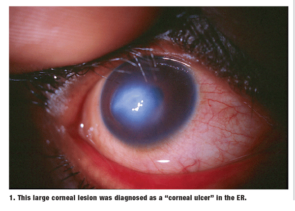
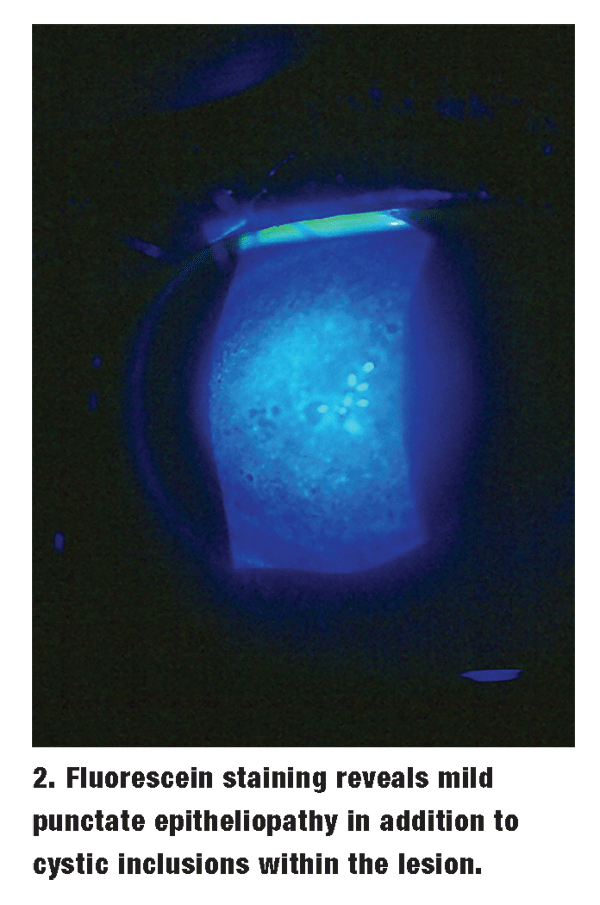
 A 19-year-old black male presents with unilateral discomfort, photophobia, decreased vision and a two-week history of a grayish corneal lesion in his right eye. Several days earlier, he had been evaluated at the local emergency room and was told that he had a corneal ulcer. He was placed on a regimen of ciprofloxacin 0.3% ophthalmic solution every two hours while awake and told to see an eye doctor the following day. He reports mild improvement in comfort since using the drops, but there is minimal change in the appearance of the eye.
His examination shows decreased vision not only in the right eye, but also in the left; pinhole occlusion reveals 20/60 O.D. and 20/30 O.S.
Biomicroscopy of the right eye reveals a well-circumscribed, bluish-white, disciform corneal defect (figure 1). Closer inspection shows that the area is grossly elevated, with cystic inclusions and dense corneal edema. There is no evidence of leukocytic infiltration or epithelial erosion. Sodium fluorescein staining shows only mild punctate epitheliopathy over the lesion (figure 2). Clearly, this is not microbial keratitis, as the patient was told in the emergency room; rather, the findings suggest corneal hydrops.

Inspection of the fellow eye reveals a steep corneal apex with superior thinning and protrusion of the lower lid upon downgaze. A quick retinoscopy shows scissoring of the reflex in the left eye.
The diagnosis now becomes clear: Unbeknownst to the patient, he has advanced keratoconus.

Keratoconus is a non-inflammatory, progressive, irreversible degeneration of the cornea that usually first manifests around puberty. It presents with a bulging forward of the inferior-central aspect of the cornea. Patients commonly report blurring and distortion of vision that gradually worsens over time.
Other common symptoms include glare and photophobia, monocular diplopia and, in some cases, ocular discomfort. The rate of progression and severity varies from patient to patient. Keratoconus is usually bilateral, but asymmetry is common.1,2
There are several classic clinical findings in keratoconus. The best-known of these is Munsons sign, a focal protrusion of the lid margin that is seen in downgaze and which corresponds to the corneal cone.
Another well-known sign of keratoconus is a scissors reflex upon retinoscopy. Slit lamp examination often reveals an annular ring of hemosiderin pigment at the cones base, referred to as Fleischers ring.
Another classic finding is Vogts striae, vertical striae in the posterior stroma that disappear with the application of digital pressure. It is also common to see an abnormal red reflex, in which a dark annular shadow surrounds the bright reflex of the cone.
Other possible biomicroscopic findings include enlarged corneal nerves, clear spaces in the anterior stroma and fine subepithelial fibrillary lines.3
While keratoconus tends to be easily recognized by most clinicians, patients may still remain undiagnosed for many years. We have seen relatively asymptomatic patients in their 20sand sometimes their 30swho were entirely unaware of their condition. These patients often demonstrate reduced acuity, but because of adaptation and high blur tolerance, they do not seek vision care. This was the case with our emergency patient; he simply had never undergone a comprehensive eye examination.
Beat the Bulge
Keratoconus results in progressive corneal ectasia and irregular astigmatism. So, gas permeable contact lenses are often employed to stabilize the condition and maximize visual acuity. As many as 75% of keratoconus patients can be maintained safely in contact lenses for their condition.4
Because of the irregular shape, conventional lens designs may not be sufficient; instead, specialized lenses attempt to vault the paracentral area while providing the necessary bearing over the cone apex. Some well-known lens designs include the Soper, McGuire, NiCone and Rose K systems.4,5
Corneal hydrops is a significant complication of advanced keratoconus. Hydrops represents an acute state of stromal edema that results from ruptures in Descemet"s membrane. The condition presents with reduced acuity, discomfort, redness and photophobia. Although corneal hydrops is self-limiting, it can result in corneal scarring.6,7
Treatment for corneal hydrops is directed at relieving the discomfort and diminishing corneal edema. Cycloplegia (e.g., 0.25% scopolamine b.i.d.) often helps alleviate the photophobia and pain associated with hydrops. Therapeutic bandage contact lenses are somewhat controversial; they may promote epithelial healing, but they also can exacerbate corneal edema. Topical hyperosmotic agents, such as 5% sodium chloride ointment, may also be used; however, these have their primary effect in the epithelium, and do little to reduce stromal edema. Topical nonsteroidal anti-inflammatory drugs (NSAIDs), such as Acular LS (ketorolac tromethamine, Allergan), or corticosteroids, such as Pred Forte (prednisolone acetate, Allergan), may be used to manage severe discomfort, although this is rarely necessary.
Corneal hydrops resolves within a few weeks to several months with supportive therapy. Practitioners should follow these patients every one to two weeks until complete resolution is noted.
Managing keratoconus can be extremely challenging. This is especially true for those late-blooming patients with keratoconus, who may be incredulous that they have a progressive, potentially sight-threatening disorder. Proper education is the key to helping these patients cope with their condition; once they understand and accept the diagnosis, appropriate therapy can be implemented.
Unfortunately, our patient was not receptive to the explanation we offered. Although we initiated treatment for his condition, he elected not to return for follow-up care.
1. Rabinowitz YS,
2. Rabinowitz YS. Keratoconus. Surv Ophthalmol 1998 Jan-Feb; 42(4):297-319.
3. Krachmer JH, Feder RS, Belin MW. Keratoconus and related noninflammatory corneal thinning disorders. Surv Ophthalmol 1984 Jan-Feb;28(4):293-322.
4. Lembach RG. Use of contact lenses for management of keratoconus. Ophthalmol Clin North Am 2003 Sep;16(3):383-94, vi.
5. Betts A, Mitchell GL, Zadnick K. Visual performance and comfort with the Rose K lens for keratoconus. Optom Vis Sci 2002 Aug;79(8):493-501.
6. Feder RS. Noninflammatory ectactic disorders. In: Krachmer JH, Mannis MJ,
7. Maguire LJ. Ectatic corneal degenerations. In: Kaufman HE, Barron BA,
Cornea, 2nd Edition.

