 History
A 57-year-old black female was admitted to the hospital after being struck by an automobile. The intensive care unit requested an ocular consult, which was prompted by marked swelling of her right eye that had persisted for three days.
Her chart revealed the presence of an orbital floor fracture without dislocation or apparent entrapment. The patient explained that she could not see because she was unable to open her eye. However, she did not complain of exceptional pain, and reported discomfort only upon upward gaze.
Her systemic history was significant for hypertension, for which she was properly medicated. Her ocular history was significant for bilateral cataracts. She had no known allergies.
Diagnostic Data
Her best-corrected visual acuity measured 20/20 O.U. at distance and near. External examination revealed ecchymosis of the right eye, with a palpable soft edema and painful area on the inferior temporal orbital rim.
There was no evidence of afferent pupillary defect or visual field involvement. The anterior segment findings were normal. Her intraocular pressure measured 14mm Hg O.U. Dilated fundoscopy was within normal limits O.U.
Your Diagnosis
How would you approach this case? Does this patient require any additional tests? What is your diagnosis? How would you manage this patient? What’s the likely prognosis?
Discussion
Additional testing included a study of ocular motility to confirm the full range of motion. Sensitivity testing was also explored to rule out the possibility of damaged sensory nerves.
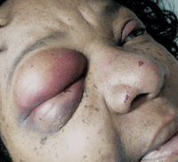
External examination of our patient showed evidence of substantial bruising.
The diagnosis in this case is inferior orbital rim fracture with orbital emphysema O.D.
Blunt trauma to the orbital rim is a frequent cause of orbital floor and medial orbital wall fractures.1-3 The specific term “blowout fracture” is reserved to connote an isolated orbital floor or medial wall fracture in the context of an intact orbital rim.1-4 Patients usually present with a history of blunt-force trauma (such as being struck with a projectile, bat or fist) or a collision injury (such as that caused by the impact of an automotive air bag or secondary to contact with an object following a fall).5
Common clinical findings include: photophobia; lacrimation associated with the post-traumatic uveal inflammation (iritis or iridocyclitis); variable facial swelling secondary to fluid or air (orbital emphysema); crepitus (a crackling noise audible when air-infiltrated tissue is palpated); gaze-evoked diplopia; and pain upon eye movement.6 Other associated collateral injuries include subconjunctival hemorrhage, ruptured globe, corneal abrasion, conjunctival laceration, hyphema, iridodialysis, lenticular subluxation, retinal detachment, vitreous hemorrhage, choroidal rupture and optic nerve evulsion. If the eye settles inferiorly or medially into the exposed sinus, enophthalmos with restricted ocular motility will be present––with or without loss of facial sensation.
The seven bones of the orbit include the frontal, zygomatic, maxillary, ethmoid, sphenoid, lacrimal and pterygopalatine.7 The other critical anatomical components are the surrounding paranasal air sinuses.8 Unfortunately, these structures leave the superior, medial and inferior walls of the orbit unsupported and vulnerable to catastrophic failure (blowout fracture, trap-door fracture) via blunt-force trauma. The sinuses surrounding the orbit include the ethmoidal air cells (anterior, middle and posterior), the sphenoidal sinuses, the maxillary sinuses and the frontal sinuses.7,8
There is some debate about the mechanism of blowout fracture. When a blunt force impacts the face, it may produce a combination of effects:
- The force may be transmitted to the eyeball. This causes the globe to strike one of the orbital walls, which results in a fracture.
- The force may strike the bone, producing a shockwave that causes “bone buckling.”
- The force may be transmitted by the globe via the principle of fluid incompressibility. This causes generalized increased orbital content pressure, or a “hydraulic” effect, resulting in fractures.9-12
The point of breakage usually occurs along the axis of least support in an area where the tissue is weakest.9-12 Because the orbital floor is not parallel to the horizontal plane, the vector of the striking force seems to affect the resultant fracture patterns.9
While all three of the aforementioned fracture mechanisms are referenced in the literature, the latter two have received increased support during the last few years.10-12 Fractures produced by the bone-buckling mechanism typically are limited to the anterior part of the orbital floor.11 In contrast, “hydraulic” fractures are often larger, involving both the anterior and posterior parts of the floor as well as the orbit’s medial wall.11 In either case––because the orbital floor has been found to have a lower threshold for fracture than the medial wall and other orbital bones––when it gives way, the globe and its attached components become unsupported, slipping below into the vacant sinus, producing visible enophthalmos and gaze-evoked, symptomatic diplopia as well as varying degrees of extraocular muscle dysfunction and infraorbital nerve hypoesthesia.10,12
During the acute stages, the treatment of a blowout fracture centers on ocular first aid. The most challenging aspect of examining patients who have encountered facial blunt-force trauma is physically opening their eyes. Facial and orbital swelling or orbital emphysema can literally force the eye closed. Here, a Desmarres lid retractor can be used as a speculum to lift the superior lid. The thin blade can be inserted between the lids and pulled up to elevate the superior lid. The examination must proceed in a logical sequence––from adnexa to posterior pole.
You must perform a CT scan to rule out concomitant maxillofacial-orbital fracture or a ruptured globe, as well as a dilated fundus exam to rule out vitreous hemorrhage, retinal tearing and retinal detachment. Furthermore, patients with significant facial lacerations may require referral to an oculoplastic surgeon for cosmetic considerations and/or to restore functional lid closure. Additionally, conduct a Seidel test to rule out perforating injuries.
Appropriate therapeutic management consists of topical anti-infective ointments for all cuts and abrasions; topical anti-infective drops for any observed corneal abrasions; and topical and oral anti-inflammatory therapy for resultant ocular and facial inflammation.
Keep in mind that the treatment of blowout fractures may not be emergent. Obviously, however, in the presence of a compressive threat to the optic nerve via swelling and retrobulbar hemorrhage, an emergency referral for lateral canthotomy and orbital decompression is necessary. Typically, surgical intervention is postponed until orbital health is consistent with a good surgical environment––unless large amounts of soft tissue are incarcerated in the bony rupture.13
Traditionally, orbital floor fractures are managed via transconjunctival and subciliary incisions; however, postoperative lid malposition is a potential complication.14
Recently, some surgeons have begun to employ an endoscopic approach to orbital floor fractures, which has the benefit of a hidden incision and improved fracture visualization.14 When the orbital floor requires replacement or reconstruction, ultra-thin, porous polyethylene implants serve as durable substitutes that mimic the pertinent anatomy and prohibit the morbidity of rejection.15
We treated our patient for mild traumatic iritis with topical cyloplegia, topical anti-inflammatory agents and oral analgesics. We suggested the addition nasal decongestant spray b.i.d. to reduce the need for nose blowing as well as a stool softener to decrease the effort required for defecation. The patient experienced uncomplicated resolution during the next three weeks, and did not require surgical intervention.
We classified her as a late glaucoma suspect, and instructed her to return every six months for IOP measurement and optic nerve evaluation.
1. Lelli GJ Jr, Milite J, Maher E. Orbital floor fractures: evaluation, indications, approach and pearls from an ophthalmologist’s perspective. Facial Plast Surg. 2007 Aug;23(3):190-9.
2. Burm JS, Chung CH, Oh SJ. Pure orbital blowout fracture: new concepts and importance of medial orbital blowout fracture. Plast Reconstr Surg. 1999 Jun;103(7):1839-49.
3. Guly CM, Guly HR, Bouamra O, et al. Ocular injuries in patients with major trauma. Emerg Med J. 2006 Dec;23(12):915-7.
4. McGwin G Jr, Owsley C. Incidence of emergency department-treated eye injury in the United States. Arch Ophthalmol. 2005 May;123(5):662-6.
5. Lehto KS, Sulander PO, Tervo TM. Do motor vehicle airbags increase risk of ocular injuries in adults? Ophthalmology. 2003 Jun;110(6):1082-8.
6. García-Medina JJ, García-Medina M, Pinazo-Durán MD. Severe orbitopalpebral emphysema after nose blowing requiring emergency decompression. Eur J Ophthalmol. 2006 Mar-Apr;16(2):339-42.
7. Snell RS, Lemp MA. The orbital cavity. In: Snell RS, Lemp MA. Clinical Anatomy of the Eye, 2nd ed. Malden, Mass.: Blackwell Science; 1998:59-77.
8. Snell RS, Lemp, MA. The paranasal sinuses. In: Snell RS, Lemp MA. Clinical Anatomy of the Eye, 2nd ed. Malden, Mass.: Blackwell Science; 1998:78-89.
9. Burnstine MA. Clinical recommendations for repair of orbital facial fractures. Curr Opin Ophthalmol. 2003 Oct;14(5):236-40.
10. Nagasao T, Miyamoto J, Nagasao M, et al. The effect of striking angle on the buckling mechanism in blowout fracture. Plast Reconstr Surg. 2006 Jun;117(7):2373-80.
11. Ahmad F, Kirkpatrick NA, Lyne J, et al. Buckling and hydraulic mechanisms in orbital blowout fractures: fact or fiction? J Craniofac Surg. 2006 May;17(3):438-41.
12. Rhee JS, Kilde J, Yoganadan N, et al. Orbital blowout fractures: experimental evidence for the pure hydraulic theory. Arch Facial Plast Surg. 2002 Apr-Jun;4(2):98-101.
13. Harris GJ. Orbital blow-out fractures: surgical timing and technique. Eye (Lond). 2006 Oct;20(10):1207-12.
14. Farwell DG, Strong EB. Endoscopic repair of orbital floor fractures. Facial Plast Surg Clin North Am. 2006 Feb;14(1):11-6.
15. Ozturk S, Sengezer M, Isik S, et al. Long-term outcomes of ultra-thin porous polyethylene implants used for reconstruction of orbital floor defects. J Craniofac Surg. 2005 Nov;16(6):973-7.
