 A 58-year-old white male who presented for a contact lens examination complained of seeing subtle spots before both eyes for the previous two weeks. His ocular history is negative for any surgery, trauma or disease.
His medical history is significant for mild hypertension and hyperlipidemia, which he controls with lisinopril 10mg q.d. and atorvastatin 10mg q.d., respectively, and benign prostatic hypertrophy, for which he takes saw palmetto. His family history is positive for heart disease. Additional medications include baby aspirin, vitamin C and vitamin E.
Because of his cardiac risk factors, the patient has annual physical examinations. At his most recent physical, his blood pressure measured 130/78mm Hg, and his total cholesterol measured 164mg/dL. High- and low-density lipoproteins (HDL and LDL) measured 51mg/dL and 97mg/dL, respectively. His stress echocardiogram testing was normal as of two years ago.
The patients visual acuity was 20/20 O.U. at distance and near. Confrontation visual fields were full to careful finger counting O.U. His pupils were equally round and reactive, and there was no afferent pupillary defect.
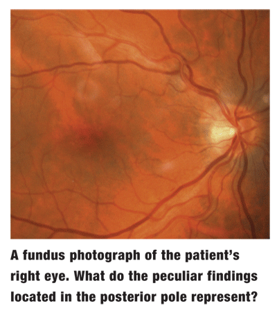
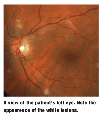
Extraocular motility testing was normal, and the anterior segment was unremarkable. Intraocular pressure measured 16mm Hg O.U. The patients fundus findings are shown in the photographs.
Take the Retina Quiz
1. What retinal findings are evident in this patient?
a. Bilateral active toxoplasmosis.
b. Diffuse cotton-wool spots.
c. Early cytomegalovirus (CMV) retinopathy.
d. Purtschers retinopathy.
2. What do the fundus findings demonstrate?
a. Focal accumulations of white blood cells.
b. Retina edema within the capillary beds.
c. Axoplasmic stasis within the nerve fiber layer.
d. Active retinitis.
3. What is the appropriate initial management for this patient?
a. Medical work-up, including complete blood count (CBC) and fasting blood glucose.
b. In-office blood pressure check.
c. No work-up at this time; follow up in six weeks.
d. Both a and b.
4. Which of these is the least likely cause of this finding in any patient?
a. Thyroid eye disease.
b. Chronic anemia.
c. A history of prior head or neck radiation.
d. A cardiac myxoma.
For answers, see below.
Discussion
Our patient has multiple cotton-wool spots (CWS), as suggested by the changes demonstrated in the fundus photographs. These result from a disruption of axoplasmic flow due to local ischemia within the retinal nerve fiber layer. On clinical exam, they appear as fluffy, white lesions within the superficial retinal layers. Patients may report small scotomas corresponding with the physical location of the CWS. The retina also appears slightly thickened due to swelling caused by the localized ischemia.
Poorly controlled hypertension and diabetes are the most common diseases associated with CWS.1 Other ischemic sources may include vaso-occlusive diseases, such as carotid stenosis, retinal vascular occlusion or hypercoaguable states.
Endogenous emboli may accompany CWS. These may be secondary to abnormalities within the carotid artery or the cardiac valves. Similarly, exogenous emboli introduced to the bloodstream may cause microvascular infarctions. Consider the possibility of collagen vascular diseases, such as systemic lupus erythematosis, any time you suspect systemic vasculitis.
Adverse responses to medical therapies, including interferon and certain forms of chemotherapy, may also manifest with CWS. Radiation therapy to the head or neck may result in either microvascular anomalies or the development of CWS during subsequent years.
Significant physical or ocular trauma may also result in CWS, presenting as Purtschers retinopathy. Finally, a disseminated bacterial or fungal infection may occur in an eye with CWS.
So, how can we treat this patient? Should we assume that the spots are the result of his systemic hypertension, or are they an insignificant finding? A paper published in 1985 looked at these questions.2 The papers authors investigated 25 consecutive patients who presented with single or multiple CWS to determine if the condition had a systemic cause. Known diabetic patients were excluded from the study.
The authors found that five patients had undiagnosed diabetes, and another five had undiagnosed systemic hypertension. Several patients had severe carotid artery obstruction, systemic lupus erythromtosis, leukemia, AIDS and metastatic carcinoma.2 Only in one case did a medical workup fail to reveal an underlying cause of CWS.
So, the discovery of any CWS merits additional medical investigation, even in an otherwise healthy patient.3 Consider that CWS may be caused by a wide variety of factors, each with serious systemic significance. A thorough work-up is warranted, including blood pressure measurement, complete blood count with differential, a blood chemistry panel and a fasting blood sugar test.
Additional studies may be warranted, including carotid and cardiac studies, erythrocyte sedimentation rate in those at risk for temporal arteritis, a rheumatoid factor test, an analysis of antinuclear antibody levels if you suspect collagen vascular disease, and a hemoglobin electrophoresis test to rule out sickle cell anemia.
We do not believe that our patients CWS resulted from his hypertension, as his blood pressure was well controlled. Further laboratory testing found grossly elevated levels of homocysteine. His level was 91mol/L, while the normal range is 0 to 13mol/L.
Homocysteine is an intermediate product derived when methionine is metabolized. When a persons normal metabolic rate is disturbed, homocysteine levels increase, a condition known as hyperhomocysteinemia.
It is not yet fully understood how hyperhomocysteinemia may accelerate atherogenesis and thrombosis, but its effects seem to be synergistic with other risk factors, such as smoking and hypertension. Hyperhomocysteinemia is a known risk factor for atherosclerotic events, including myocardial infarction, cerebrovascular infarction and carotid artery disease.4,5
Reports link hyperhomocysteinemia to retinal artery and vein occlusions, ischemic optic neuropathy, exudative (wet) age-related macular degeneration and pseudoexfoliation glaucoma.6-10 Moderate hyperhomocysteinemia may be caused by deficient intake of B vitamins and/or folic acid.
We put our patient on vitamin B and folic acid supplements. He responded very well and demonstrated no deterioration in either ocular or cardiac status. On follow-up, the CWS resolved on its own without complications. After six months of nutritional supplementation, his homocysteine levels improved from 91mol/L to 25
mol/L. Additionally, he no longer had any ocular signs or visual symptoms.
Thanks to Christian P. Guier, O.D., at the Mayo Clinic in
Retina Quiz answers: 1) b; 2) c; 3) d; 4) a.
1. Arroy JG. Cotton-wool spots may challenge diagnosis. Rev Ophthalmol 2004 Apr;11(4):111-5.
2. Brown GC, Brown MM, Hiller T, et al. Cotton-wool spots. Retina 1985 Fall-Winter;5(4)206-14.
3. Remky A, Arend O. Single isolated cotton-wool spots. Ophthalmologica 2000; 214(2):143-8.
4. Haynes WG. Hyperhomocysteinemia, Vascular function and atherosclerosis: effects of vitamins. Cardiovasc Drugs Ther 2002 Sep;16(5):391-9.
5. Miller A, Mujumdar V, Shek E, et al. Hyperhomocysteinemia induces multiorgan damage. Heart Vessels 2000;15(3):135-43.
6. Vine AK. Hyperhomocysteinemia: A risk factor for central retinal vein occlusion. Am J Ophthalmol 2000 May;129(5):640-4.
7. McGumpsey SJ, Woodside JV, Bamford L, et al. Retinal vein oclusion, homocysteine, and methylene tetrahydrofolate reductase genotype. Invest Ophthalmol Vis Sci 2005 Dec;46(12):4712-6.
8. Di Crechio L, Parodi MB, Sanguinetti G, et al. Hyperhomocysteinemia and the methylenetetrahydrofolate reductase 677C-T mutation in patients under 50 years of age affected by central retinal vein occlusion. Ophthalmology 2004 May;111(5):940-5.
9. Stanger O, Weger M, Obeid R, et al. Impairment of homocysteine metabolism in patients with retinal vascular occlusion and non-arteritic ischemic optic neuropathy. Clin Chem Lab Med 2005;43(10):1020-5.
10. Axer-Seigel R, Bourla D, Ehrlich R, et al. Association of neovascular age-related macular degeneration and hyperhomocysteinemia. Am J Ophthalmol 2004 Jan;137(1):84-9.
