 The CDC recently delivered some heartening news: Cholesterol levels among U.S. adults have dropped an average of 10 points over the past two decades.1 However, an estimated 71 million American adults have high cholesterol, so there is still a ways to go.2
High cholesterol increases your patients’ risk for coronary heart disease, heart attack and stroke.3 Its damaging effects are not limited to arteries and vessels, as high cholesterol can significantly affect vision and eye health as well.
This month, we’ll review the basics about cholesterol, its relationship with ocular health and common treatments.
Back to Basics
Cholesterol, comprised of a lipid and a sterol, is so essential for survival that the body makes about 75% of its supply—with the remainder coming from the foods that we eat.3 The body needs cholesterol to produce the outer membrane of cells, create the bile acids that help digest food in the intestine and make vitamin D and hormones, like estrogen and testosterone.4
Because lipids are oil-based and blood is water-based, it isn’t easy for cholesterol to flow through the bloodstream.5 For example, if cholesterol were dropped directly into the bloodstream, it would form into blobs like oil does in vinegar and would be unusable.
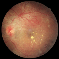
This image highlights fat (exudate) deposits in the retina of a diabetic patient.
To allow it to mix more easily with blood, the body packages cholesterol and other fats into small protein-covered particles called lipoproteins (lipid + protein).5
The five major types of lipoproteins are:6
- Chylomicrons.
- Very low-density lipoproteins (VLDL).
- Intermediate-density lipoproteins (IDL).
- Low-density lipoproteins (LDL).
- High-density lipoproteins (HDL).
The proteins that combine with the cholesterol are called apolipoproteins. The fat in these particles is comprised of cholesterol, triglycerides and phospholipids (which help to hold the whole particle together).5 The body needs triglycerides for energy but, like cholesterol, too much is bad for the arteries and the heart.5,7
Testing and Causative Factors
Because there are no symptoms of high cholesterol, it is crucial that patients keep up with regular monitoring through simple blood testing that can provide a breakdown of cholesterol levels. Elective determinations of plasma lipid concentrations should be made after an overnight fast (preferably 10 to 14 hours). Directly measured components include triglycerides, total LDL and HDL.
High cholesterol can result from a number of possible causes: a genetic predisposition, a diet too high in cholesterol or the inability to excrete cholesterol efficiently.5 Some of these factors are unavoidable but treatable, while others are entirely controllable.
For instance, evidence suggests that Americans tend to have higher blood cholesterol levels than people in the Far East or Africa largely because of America’s high-fat, high-cholesterol diet.5,8
Patients with
abnormally high LDL cholesterol levels or uncharacteristically low HDL
cholesterol levels can be at risk for a number of ocular comorbidities.
Cholesterol and the Eye
How Cholesterol Moves
After a meal, the intestines absorb the nutrients the body needs from food. In the small intestine, intestinal enzymes degrade lipids into component fatty acids. They are then reassembled and bundled into new triglyceride molecules and packaged, along with some cholesterol, into chylomicrons.5,7,9
At the same time, carbohydrates and proteins pass to the liver, which converts some of these nutrients to triglyceride molecules, packages them with apolipoproteins and cholesterol, and releases them into the blood as VLDL.7
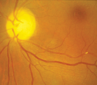
In this patient, you can see cholesterol plaque has lodged in a retinal artery.
As mentioned earlier, the body produces the majority of cholesterol. Even if an individual were to eat a completely cholesterol-free diet, the body would make about 1,000mg of cholesterol—the amount it needs to function properly.5 The body can regulate the amount of cholesterol in the blood, creating more when diet doesn’t provide enough.5
Nearly all cells in the body can make the cholesterol they need, but the liver is especially efficient in producing cholesterol, which makes it central to the regulation of cholesterol levels.5,6,9
The liver packages most of its cholesterol into lipoproteins that are delivered to cells throughout the body, which provides each cell with an additional supplement to what it makes on its own.5 Once released into the circulation, VLDLs supply free fatty acids to tissues as well as transport fats. When they give up their fat, VLDLs turn into IDLs.
Over time, IDLs turn into LDL cholesterol. If there are too many LDL particles in the bloodstream, they deposit the cholesterol onto the walls of the arteries and blood vessels, which can lead to build-up and blockages.5 The liver and the intestines also make HDLs, which scavenge cholesterol from the blood and artery walls and take it to the liver for disposal.5,7,9
Cholesterol Medications
Intervention studies in the 1990s showed that cholesterol reduction by means of diet or drugs reduced the risk of development or progression of coronary heart disease.6
If an individual has high cholesterol, a total cholesterol level that is 200mg/dL or higher or a low-density lipoprotein cholesterol (LDL) level that is 130 mg/dL or higher, a cholesterol-lowering medication may be recommend (depending upon other risk factors involved).10
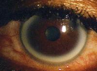
Corneal arcus is a particularly sensitive sign of familial hypercholesterolemia. Photo: Howell Findley, O.D.
Let’s look at some of the most commonly used drugs:4,7
- Statins (e.g., Lipitor [atorvastatin, Pfizer]) are the most widely prescribed cholesterol-lowering drugs. They block HMG-CoA reductase, a key liver enzyme involved in the production of cholesterol.
- Bile acid sequestrants (e.g., Colestid [colestipol, Pfizer]) inhibit the body’s absorption of dietary cholesterol.
- Niacin (nicotinic acid) is said to increase HDL levels and decrease triglyceride and LDL levels when used at high doses.
- Omega-3 fatty acids increase the level of HDL and lower the level of trigylcerides.
- Cholesterol absorption inhibitors (e.g., Zetia [ezetimibe, Merck]) decrease the amount of cholesterol absorbed from food in the digestive tract.
- Fibrates (e.g., Lopid [gemfibrozil, Pfizer]) decrease triglycerides by reducing the liver’s production of VLDL and accelerating its removal from the blood.
Most cholesterol medications lower cholesterol with few side effects, but effectiveness may vary from person to person.
Healthy lifestyle choices—such as smoking cessation, exercise, weight loss, stress management, and a healthy diet that is low in saturated fat, cholesterol and salt—should be considered.5
1. Carroll MD, Kit BK, Lacher DA, et al. Trends in lipids and lipoproteins in US adults, 1988-2010. JAMA. 2012 Oct 17;308(15):1545-54.
2. CDC. Vital signs: prevalence, treatment, and control of high levels of low-density lipoprotein cholesterol. United States, 1999–2002 and 2005–2008. MMWR. 2011;60(4):109-14.
3. American Heart Association. Cholesterol. 2012 Aug 8. Available at:
www.heart.org. Accessed November 7, 2012.
4. Understanding cholesterol and heart health. NIH Medline Plus. 2012;7(2):4.
5. Freeman MW, Junge C. The Harvard Medical School guide to lowering your cholesterol. Boston: McGraw-Hill; 2005:1-12.
6. Lange RA, Hilis LD. Coronary heart disease. In: Andreoli and Carpenter’s Cecil Essentials of Medicine. 7th ed. Philadelphia: Elsevier Inc.; 2007:97-117.
7. What to do about high cholesterol: a Harvard Medical School special health report. Boston: Harvard Health Publications; 2012.
8. All in the family: when high blood cholesterol occurs in families. NIH Medline Plus. 2012;7(2):8-9.
9. Linder T, Melby AE. Cholesterol, lipoproteins, and the liver. University of Washington. Available at:
http://courses.washington.edu/conj/bess/cholesterol/liver.html. Accessed November 7, 2012.
10. Pyeritz RE. Disorders of lipid metabolism. In: Andreoli and Carpenter’s Cecil Essentials of Medicine. 7th ed. Philadelphia: Elsevier Inc.; 2007:617-24.
11. Miller M. Signs and symptoms of dyslipidemia: ophthalmologic signs. Medscape Education. Available at:
www.medscape.org/viewarticle/585045. Accessed November 7, 2012.
12. Fernández A, Sorokin A, Thompson PD. Corneal arcus as coronary artery disease risk factor. Atherosclerosis. 2007 Aug;193(2):235-40.
13. Patterson L. Arcus senilis: an important forensic physical finding. Am J Forensic Med Pathol. 1982;3(2):115-8.
14. Park YH, Lee YC. Images in clinical medicine. Lipemia retinalis associated with secondary hyperlipidemia. N Engl J Med. 2007 Sep 6;357(10):e11.
15. O’Mahoney PR, Wong DT, Ray JG. Retinal vein occlusion and traditional risk factors for atherosclerosis. Arch Ophthalmol. 2008 May;126(5):692-9.
16. Kohnle D. Xanthelasma and xanthoma. Beth Israel Deaconess Medical Center. Available at:
www.bidmc.org/YourHealth/ConditionsAZ.aspx?ChunkID=202823. Accessed November 7, 2012.
