
Q: I have a patient who recently experienced a significant, full thickness/large abrasion after being hit by a tree branch. She underwent LASIK surgery six months ago. How do you balance the concerns for the use of steroids to minimize and manage the diffuse lamellar keratitis (DLK) after such an injury vs. the potential for fungal infection? Also, since this injury is hallmarked by pain and iridocyclitis, how do you differentiate between DLK and infection?
A: If a dense infiltrate is not present, which is inherent to infectious keratitis (both fungal and bacterial) and not to DLK, the patient likely does not have fungal keratitis, says optometrist Paul Karpecki, of Kansas City, Mo. I have found this to be true even when a patient, such as this one, has sustained a corneal abrasion as the result of contact with vegetable matter, he says. Also, research has shown that fungal keratitis in the United States is extremely rare.1
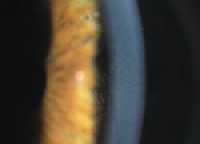 |
| DLK presents with accumulations of white blood cells at the interface and no mucopurulent discharge. Also, the infiltrate in DLK is often more peripheral and becomes central over time. |
Ophthalmologist Thomas S. Boland, of Scranton Pa., says the risk of this patient developing DLK is significantly higher than the risk of her developing fungal keratitis because she underwent LASIK fairly recentlymaking her more susceptible to DLK. Treat this patient with a mild steroid, such as FML (fluorometholone alcohol, Allergan) q.i.d. until the cornea completely reepithelializes. This generally takes about four days, he says. Then, monitor her for signs of fungal keratitis.
This patient should also be treated prophylactically with an antibiotic drop, such as Zymar or Vigamox (moxifloxacin HCl ophthalmic solution, Alcon) q.i.d. until the eye is completely reepithelialized, Dr. Boland says. Have the patient do this in addition to the steroid drops to prevent the onset of bacterial keratitis, he says.
So, how do you differentiate between DLK and infection since the injury described above is hallmarked by pain and iridocyclitis? The aforementioned presence of a focused infiltrate, which is inherent to infectious keratitis, and accumulations of white blood cells at the interface, which are inherent to DLK, is one way to differentiate between DLK and infection, Dr. Karpecki says. In addition, monitor the patient closely for signs and symptoms of infection, including increased pain and anterior chamber reaction.
Other differentiating characteristics: The infiltrate in DLK is often more peripheral and becomes central over time. With infectious keratitis, the infiltrate is located where the abrasion occurred, which is usually central, Dr. Karpecki says. Also, DLK is not accompanied by mucopurulent discharge. Infectious keratitis is.
1. Srinivasan M. Fungal keratitis. Curr Opin Ophthalmol 2004 Aug;15(4):321-7.
2. Rosa RH Jr, Miller D, Alfonso EC.The changing spectrum of fungal keratitis in south Florida. Ophthalmology. 1994 Jun;101(6):1005-13.
