 A 54-year-old Hispanic male presented for his annual eye exam. He wore progressive lenses and did not report any vision problems.
Approximately four months earlier, the patient had been hospitalized for cryptococcal meningitis. At that time, he was in the hospital for approximately three weeks. He reported no ocular sequelae from the meningitis.
His systemic history was positive for HIV, and the patient wanted to make certain that there was nothing wrong with his eyes. His current medications included Diflucan (fluconazole, Pfizer), Celexa (citalopram hydrobromide, Forest Pharmaceuticals), Imitrex (sumatriptan, GlaxoSmithKline), Abilify (aripiprazole, Bristol-Myers Squibb) and clonazepam.
On examination, his best-corrected visual acuity measured 20/20 O.U. Confrontation fields were full to careful finger counting O.U. His pupils were equally round and reactive, with no evidence of afferent defect. The anterior segment examination was unremarkable in each eye. Intraocular pressure measured 12mm Hg O.U.
Dilated fundus exam showed a clear vitreous O.U. Both optic nerves appeared healthy, with small cups with good rim coloration and perfusion.
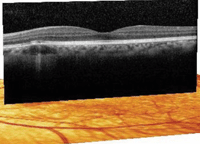
The macula and periphery of the right eye were normal. The peripheral retina in the left eye was normal; however, we noted a small, yellow-white lesion located between the optic nerve and macula (figure 1). We obtained a spectral domain optical coherence tomography (SD-OCT) scan of the left eye to further evaluate the lesion (figure 2).
Take the Retina Quiz
1. How would you describe the appearance of the lesion located in our patient’s left eye?
a. Nonspecific depigmentation of the retinal pigment epithelium (RPE).
b. Area of active retinitis.
c. Area of active choroiditis.
d. Both a and c.
2. What does the SD-OCT scan show?

1. A view of our patient’s left posterior pole. Note the yellow-white lesion located between the optic nerve and macula.
a. Normal retina.
b. Neurosensory detachment.
c. RPE detachment.
d. Localized choroidal lesion.
3. What is the likely diagnosis?
a. Nonspecific depigmentation of the RPE.
b. HIV retinopathy.
c. Cytomegalovirus retinitis.
d. Cryptococcal choroiditis.
4. How should we manage this patient?
a. Observation.
b. Referral to infectious disease specialist.
c. Antiviral therapy.
d. Antibiotic therapy.
For answers, scroll to the bottom of the page.
Discussion
At first glance, the retinal lesion located in the left eye looked like nothing more than a nonspecific RPE depigmentation. However, we had just seen the patient a year earlier and did not notice the lesion. Given his recent medical history, we ordered the SD-OCT scan. And, without question, we were surprised at the results.
The SD-OCT revealed extensive retinal and choroidal detail. Within the choroid, we observed a localized circular lesion that was located below the area of RPE depigmentation. It stood out as being optically empty when compared to the surrounding, more homogenous choroid.
Above the lesion, the RPE appeared slightly elevated and the photoreceptor integrity layer was disrupted marginally. Had this presentation occurred in the fovea, his vision likely would have been affected. But, because it was located outside the fovea, he was asymptomatic.

2. Here is the SD-OCT scan of our patient’s left eye. What does it reveal?
Having recently been hospitalized for cryptococcal meningitis, the retinal lesion was highly suspicious for choroidal infiltration. Cryptococcal meningitis is caused by the fungus Cryptococcus neoformans, which is found in various soils and plants from around the world.1
Most infections caused by C. neoformans affect only the lungs; however, meningitis may result in patients with weakened immune systems secondary to HIV, diabetes and lymphoma.1 And indeed, our patient’s CD4 count was 268, which indicated that he was
sufficiently immuncompromised to develop cryptococcal meningitis.
Ocular involvement from cryptococcal meningitis presents in approximately 6% of patients.2 In addition to choroiditis, other ocular complications include chorioretinitis, vitritis, endophthalmitis and neuroretinitis. Further adverse complications also may arise from the meningitis, such as papilledema, ophthalmoplegia, ptosis, optic atrophy and sixth nerve palsy.2
Fortunately, it appeared that our patient’s infection was limited to a small area located within his choroid. Of interest, he was already taking a medication for his crytococcal meningitis––Diflucan. This raises two important questions:
- Was the infection much worse several months ago, when the patient was first hospitalized with the meningitis?
- If the presentation was new, was the antifungal medication being dosed at an adequate therapeutic level to treat the choroiditis?
Given that this lesion was isolated, and he didn’t have any other ocular complications, we elected simply to observe our patient. We sent a note to his infectious disease physician to inform him of our findings. Also, we asked our patient to continue use of his antifungal medications.
We saw the patient several times during the subsequent five months and documented slow resolution of the RPE depigmentation and the choroidal lesion. Thereafter, he continued to be asymptomatic.
1. Kauffman CA. Cryptococcosis. In: Goldman L, Ausiello D (eds.). Cecil Medicine. 23rd ed. Philadelphia: Saunders-Elsevier; 2007:357.
2. Carney MD, Combs JL, Waschler W. Cryptococcal choroiditis. Retina. 1990;10(1):27-32.
Answers
1. d
2. d
3. d
4. a

