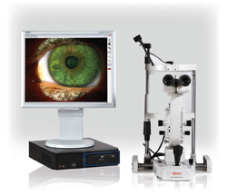
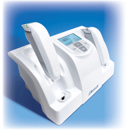
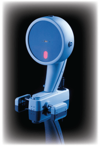
 Digital Slit Imaging System, Kowa Optimed
The Digital Slit Imaging System, from Kowa, features live video display, high-resolution capture and replay, as well as still-frame capture from live or recorded video. The user can also select a blue-free filter for enhanced viewing.
The system includes a joystick trigger for still image capture, and a dual footswitch for still image or video capture. The user can compare frame-by-frame images and keep a historical record of digital images.
Go to www.kowa-usa.com.
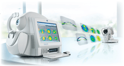
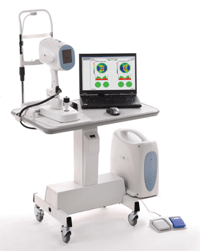
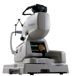 3D OCT-2000, Topcon
The 3D OCT-2000, from Topcon, incorporates a high-resolution fundus camera and user-friendly color touch screen interface and features an integrated 12.3 megapixel fundus camera.
Its proprietary FastMap software enables dynamic viewing of the OCT data, which renders 3-D, 2-D and fundus images simultaneously. These images can be quickly exported for presentations or for education. FastMap also includes noise-reduction algorithms and scanning technology for the creation of B-scan images.
The included Pin-Point Registration software indicates the location of the captured OCT image within the fundus image. The “compare” function allows the user to view serial exams in a comparison view and with different analytical tools.
The 3D OCT-2000 integrates with Topcon’s EyeRoute Image Management System to provide connectivity and access to images anywhere in the system.
Go to www.topconmedical.com.
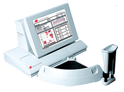 Foresee PHP, Reichert
The Foresee PHP, by Reichert, is approved for both the detection of choroidal neovascularization and the long-term monitoring of dry AMD.
“I’m a huge believer in this piece of technology,” says Kristopher May, O.D., of Coldwater, Miss. “I’ve had it save patients’ vision. A patient of mine who was in her 20s had a choroidal neovascular membrane—this device detected it when she still saw 20/15.”
The PHP performs a self-paced functional test of the 14° macular area based on hyperacuity—the patient’s sense of the relative spatial localization of multiple objects. Features of the Foresee PHP include Normative Database Analysis to aid in the early detection of CNV and quantitative automated threshold visual field test for the monitoring of AMD.
“Data storage is on flash drives, so it can be viewed on any other computer,” says Dr. May. “Also, there’s a huge amount of statistical analysis that happens, so instead of just getting the report back, the device gives you P values for the area, how much is affected, how it’s changing, etc. The analysis builds on the patient’s historical data. And it’s intuitive—it does this automatically.”
The noninvasive exam takes from three to five minutes per eye.
“For those patients who are at high risk and are defending their good eye against converting from dry AMD to wet AMD, this is a very useful tool,” says Dr. May.
Go to www.reichertoi.com/foresee.html.
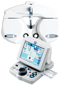 TRS-3100, Marco
In partnership with Nidek, Marco introduces the TRS-3100, which follows in the footsteps of the TRS-5100 and features a new ergonomically designed keypad and phoropter. The TRS-3100 is the first refraction system to offer a completely wireless interface with a Marco autorefractor and autolensmeter. The built-in IC Card Reader allows for seamless integration and connection, without any cable logistics.
The responsive operator keypad makes refining sphere, cylinder and prism lenses smoother and faster than manual examinations. Also, during near point tests, power convergence and power prism diopter are adjusted automatically. Prism diopter can be adjusted independently for a more accurate and reliable measurement.
The TRS-3100 offers many of the same features as the TRS-5100. With the IC Card Reader and Marco Connect (Marco’s proprietary product interface software, provided to all EMR companies), the practitioner can transfer data from pre-test equipment to the TRS-3100 and download the information to an EMR system.
Go to www.marco.com.
 TearLab Osmolarity System, TearLab
The TearLab Osmolarity System enables management of patients with dry eye disease via quantitative, objective endpoints. The entire technician-administered process requires less than 30 seconds from sample to result.
The TearLab requires 50nL of tear fluid for the test. It measures osmolarity in the human tears to aid in the diagnosis of dry eye disease. The test utilizes a temperature-corrected impedance measurement to provide an indirect assessment of osmolarity. And, through the application of a lot-specific calibration curve, osmolarity is displayed as a numeric value, which helps you discuss this finding with your patients, says the company.
“The TearLab provides a strong, objective measurement of tears,” says Dr. May. “It’s positioned to take advantage of the market.”
Go to www.tearlab.com.
 Visante Omni, Carl Zeiss Meditec
The Visante Omni, by Zeiss, integrates anterior topography from the company’s Atlas cornea topographer with Visante OCT pachymetry.
What links the two is the company’s V-Trac Registration System, which enables Visante Omni to generate posterior topography through corneal vertex alignment. The system utilizes a series of strict criteria to prevent potential misalignment.
The Visante Omni produces high-resolution images, such as full-width anterior segment imagery and complete anterior chamber angle visualization and measurement, and it creates a Holladay Report (a one-page overview of the patient’s corneal pachymetry and topography data) for advanced analysis.
Go to www.meditec.zeiss.com/us.
 Spectralis, Heidelberg Engineering
Heidelberg Engineering’s Spectralis family includes five different models: OCT, OCTPlus, HRA, FA+OCT and HRA+OCT. Each device includes different features that are specifically tailored to the needs of various practices. All include TruTrack Image Alignment Technology, which enables the user to accurately document progression over time. TruTrack places follow-up scans at precisely the same location as previous scans, enabling the detection of subtle change. It also includes dynamic selection of reference images for progression analysis, manual adjustment of the layer segmentation lines, improved display options and expanded printing options. The software filters image noise and creates wide-field pan-retinal images in real time. The user can customize the display on the instrument and networked viewing stations, and printing options provide similar capabilities.
The HRA+OCT offers SD-OCT, FA, autofluorescence, red-free and infrared imaging modes, as well as single frame, multi-frame movie, stereo, mean, composite, real time and tomography imaging options. It takes 40,000 A-scans/second and features an axial resolution of 3.9µm and a transverse resolution of 14µm. Viewing options include color, spectrum, heat and high-frequency scales; grayscale; 2-D and 3-D modeling; thickness map; and thickness graph or progression.
Go to www.heidelbergengineering.com.
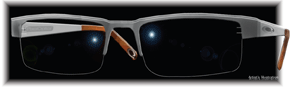 emPower!, PixelOptics
While dynamic electronic spectacle lenses aren’t diagnostic technology, they still speak to a significant segment of your patients. The emPower! brand, from PixelOptics, is intended for patients who wear bifocals or progressive lenses. These glasses include an electronically activated near focus zone. The wearer can turn this zone on and off manually, or set it to turn on automatically.
This zone is intended to provide a wide field of view and allow for reduced distortion between viewing zones. The battery is hidden in the spectacle frame, and it is rechargeable.
“The miniaturization [of technology] is very impressive,” says Dr. May. “But the real question is: Can it translate into something that patients will want to acquire? There needs to be a massive patient education movement.”
Go to www.pixeloptics.com.
 Pentacam, Oculus
The Pentacam, from Oculus, includes a rotating Scheimpflug camera and can perform a complete measurement of the anterior segment—including a precise analysis of the central cornea—in less than two seconds. It detects extraneous eye movements and corrects for them during calculation. The resulting data points serve as the basis for a three-dimensional model of the complete anterior segment.
The Pentacam’s non-contact analysis includes the entire cornea, anterior chamber and crystalline lens. It objectively determines the central radii; corneal asphericity; curvature and elevation; chamber angle, volume and elevation; and lens transparency.
Three models of the Pentacam are available: the Pentacam Basic, the Pentacam Classic and the Pentacam HR.
The Pentacam Basic includes the features an optometrist needs to monitor patients without including features that wouldn’t be used as often, says Dr. May. “The idea is to make the product more affordable—it provides extremely good data without features that the O.D. wouldn’t use.”
Go to www.oculus.de/us.
 iVue, Optovue
The iVue, by Optovue, performs retina, glaucoma and cornea scans and creates reports for each. This SD-OCT offers 5µm resolution and 26,000 A-scans per second. It also performs change and fellow-eye comparative analysis, and its ergonomic design streamlines user workflow. The user operates a touch screen and targets the patient’s eye via a live video and “en face” image.
The iVue’s imaging capabilities are broken into three groups—retina, glaucoma and cornea/anterior seg.
• Retina. The iVue has a high-resolution cross line scanner, adjustable scan rotation, fast retina mapper, change analysis and report of both eyes.
• Glaucoma. The iVue includes retinal nerve fiber layer mapping and analysis, TSNIT analysis, change analysis and report on both eyes.
• Cornea/Anterior segment. The iVue features pachymetry mapping and assessment, change analysis, high-resolution corneal pachymetry line scan and high-resolution angle imaging and measurement.
