 |
 |
Episcleritis
Episcleritis is the least symptomatic and least vision-threatening of the ocular shell conditions. Pain is typically absent or mild. Episcleritis is almost always unilateral, and onset may literally occur overnight.
The signature clinical presentation of episcleritis is sectorial injectionusually concentrated in either the nasal or temporal quadrant. Cells do not typically appear in the anterior chamber, except in the less common nodular form.
To differentiate episcleritis from scleritis and uveitis, apply 2.5% topical phenylephrine. Episcleritis will most likely blanch. You can also make a differential diagnosis by gently manipulating the affected area with a cotton-tipped applicator. Doing so distinguishes the level of inflammation. However, be sure to anesthetize the eye beforehand.
Finally, keep in mind that while true episcleritis appears spontane-ously, mechanical irritation from dry eyes and trichiasis can cause a sectorial pseudoepiscleritis. Therefore, always look for a mechanical cause.
Since the inflammation in episcleritis is superficial, virtually all steroids are acceptable for treatment. These include fluorometholone, rimexolone, loteprednol and generic prednisolone compounds. Dosing q.i.d. is usually sufficient, and cycloplegia is typically unnecessary.
Unlike episcleritis, scleritis presents a significant risk of vision loss due to collateral damage. Scleritis involves deeper ocular tissues, and is therefore more symptomatic than episcleritis. Patients characteristically report a dull, achy pain. Onset is often slow, building over several days. Hence, some scleritis patients are much less symptomatic upon initial presentation than you might anticipate. Scleritis is usually bilateral, although often asymmetric.
The common clinical presentation of scleritis is a deep red or violet hue. Involvement is usually circumferential, although sectorial injection is also possible (further confounding the differential diagnosis). Cells often appear within the anterior chamber.
The choroid, cornea, retina and even the optic nerve are subject to pathological inflammatory sequelae. Additionally, scleral thinning (scleromalacia perforans) poses the risk of global rupture.
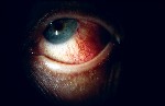 |
| Episcleritis sectorial injection. |
To differentiate scleritis from episcleritis and uveitis, view the eye under regular sunlight conditions (e.g., outside or near a window). This helps reveal the conditions true color. Topical phenylephrine does not reveal significant blanching here.
Because scleritis generally has a secondary uveitisdue to the spread of inflammation to adjacent tissuestreatment dictates topical corticosteroids and cycloplegia. Penetration is key when dealing with scleritis, so choose steroids such as Pred Forte (prednisolone acetate 1%, Allergan) or 0.5% lote-prednol. These steroids can reach the ciliary body and should be used every two to four hours, depending on the severity.
Unfortunately, topical therapy alone is not enough, so youll need to incorporate oral anti-inflammatory medications into your treatment plan. Ibuprofen (600 to 800mg q.i.d.) is a good choice. Other options include naproxen sodium (250 to 500m.g. t.i.d.), indomethacin (75mg b.i.d.) or prednisone (60 to 100mg q.d.). Recalcitrant cases may require immunomodulatory agents, such as methotrexate or cyclosporine.1,2
Uveitis
Uveitis poses a significant risk to vision. Like scleritis, uveitis typically is very symptomatic, although sometimesparticularly in older individualsthe discomfort is proportionately less than the level of inflammation suggests. Pain in uveitis may be severe, and bright lights exacerbate it.
Uveitis is unilateral in most cases. Bilateral uveitis likely indicates an underlying autoimmune or systemic disease. Onset may be sudden or gradual depending on the etiology.
Uveitis presents as a circumlimbal flush injection pattern (ie., increased vascular dilatation overlying the ciliary body). However, involvement in this area alone is rare. Uveitic eyes often exhibit diffuse deep vessel engorgement. The characteristic biomicroscopic findings include cells and flare within the anterior chamber. Other findings, which are typically absent in episcleritis and scleritis, include posterior synechiae, peripheral anterior synechiae and keratic precipitates.
These manifestations cause significant visual risks. Our biggest concern: the potential for intraocular pressure (IOP) elevation due to various mechanisms, such as inflammatory debris. This is not common in episcleritis but may occur in scleritis due to angle closure.
To differentiate uveitis from episcleritis and scleritis, instill a topical cycloplegic (e.g., 0.25% scopolamine) to see if the pain subsides. The more significant the pain, the more likely you are dealing with uveitis. The application of topical phenylephrine may cause mild blanching in uveitis, but this is not easily discernable.
Uveitis demands aggressive topical therapy with cycloplegic agents and corticosteroids. While oral anti-inflammatories may be helpful, they are rarely critical. For cycloplegia, we typically choose a powerful agent, such as 0.25% scopolamine or 1% atropine b.i.d. or t.i.d. Among corticosteroids, choose 1% Pred Forte and 0.5% loteprednol. Remember to dose consistent with the reaction. Have the patient instill steroids every 15 to 30 minutes for the first six to 12 hours in severe cases, then taper the drops to every two to four hours. Also, recognize that uveitis may persist for weeks or months (the patients response to the steroid dictates treatment length). Tapering medications in such cases can be challenging. Therefore, you must remember to monitor patients who are on long-term steroid therapy for IOP elevation and cataract formation.
Like the shell game played by street and carnival gamesters, the ocular shell game can be tricky and misleading. We must also realize that episcleritis, scleritis and uveitis may indicate underlying systemic disease, such as rheumatoid arthritis, lupus, inflammatory bowel disease, syphilis, Reiters syndrome, Behets disease, gout, Lyme disease and even giant cell arteritis.3,4 In severe or recalcitrant cases, realize that patients may require a systemic work-up.
1. Kaplan-Messas A, Barkana Y, Avni I, Neumann R. Methotrexate as a first-line corticosteroid-sparing therapy in a cohort of uveitis and scleritis. Ocul Immunol Inflamm 2003 Jun;11(2):131-9.
2. Hillenkamp J, Kersten A, Althaus C, Sundmacher R. [Cyclosporin A therapy in severe anterior scleritis. 5 severe courses without verification of associated systemic disease treated with cyclosporin A]. Ophthalmologe 2000 Dec;97(12):863-9.
3. Jabs DA, Mudun A, Dunn JP, Marsh MJ. Episcleritis and scleritis: clinical features and treatment results. Am J Ophthalmol 2000 Oct;130(4):469-76.
4. Pavesio CE, Meier FM. Systemic disorders associated with episcleritis and scleritis. Curr Opin Ophthalmol 2001 Dec;12(6):471-8.
