 The start of the congress was a delightful evening reception held in the Parliament House, and the highlight was a tradition the conference organizers called “Hypotheticals.” Hypotheticals are intended to be a lighthearted way of exploring patient management issues, rather than delving into diagnostic care.
For each case, a rotating panel of conference speakers played the role of various health care providers. The goal of these panels was to entertain the congress attendees and help them consider some of the management issues in modern optometric practice.
As one of the panelists, the aspect that I found most fascinating was that, on each panel, the “hypothetical doctors” were always an optometrist, an ophthalmologist—and a primary care physician.
I came to realize just how much the optometrists in Australia work with primary care physicians.
I informed my hosts that, in the United States, optometrists can and frequently do order lab studies and magnetic resonance imaging. That is not the case for Australian optometrists. In fact, even primary care physicians in Australia aren’t allowed to order MRIs; that remains the realm of specialists.
However, I wondered if it might be more in our patients’ best interests if, when we suspect that systemic disease may be a factor, we coordinate a management strategy with the primary care physician, share insights and clinical suspicions, and allow these medical specialists to order the appropriate testing rather than doing it ourselves. Clearly, internists have unique training and perspectives on systemic disease.
In this month’s column, I will examine the rationale of forming better working relationships with internists when encountering patients who need additional systemic evaluation.
Comanaged Therapy
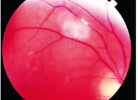
An asymptomatic patient with a
retinal embolus may not need carotid evaluation, but rather a targeted
atherosclerotic evaluation by an internist.
A recent experience showed me the value of good interpersonal relationships with primary care physicians. An internist referred a 33-year-old man for a comprehensive ophthalmic evaluation; the patient complained of headaches with visual aura. Initial blood work and neurological screening by the internist were unremarkable. The patient had perfectly normal threshold visual fields. The optic discs were normal in each eye, but there was a small juxtapapillary hemorrhage in his left eye.
I discussed the findings with the referring internist, and together we decided upon a list of differential diagnoses that included blood dyscrasias and malignancies, collagen vasculopathies, and infectious diseases. Because the presenting complaint was headache, we decided that neuroimaging should be done to rule out occult intracranial pathology, even though the visual field was normal. I could have ordered all the testing myself, but coordinating care with the internist clearly had advantages in reducing any inappropriate and unnecessary testing.
The true advantage of coordinating care with the patient’s internist came quickly and clearly. While all blood testing came back normal, the MRI indicated a massive 9.5cm tumor in the right frontal-parietal region that was causing a midline shift. The internist received a call from the radiologist late on a Friday evening. Immediately, the internist arranged for hospital admission and subsequent treatment with anti-seizure medications, steroids and acetazolamide, as well as an emergency neurosurgical consult.
Had I been the one ordering the testing, the subsequent follow-up, hospital admission, and emergency neurosurgical consultation would have been inherently more difficult.
In another recent scenario, an elderly patient presented asymptomatically, but was found to have a retinal embolus—an indication of significant systemic vascular disease. The most common associated systemic diseases are hypertension, cardiovascular disease and stroke.1-7 In many studies, the prevailing risk factor for retinal emboli is smoking.1-4
The presence of asymptomatic retinal emboli indicates risk for future embolic events, so a preventative approach dictates that all modifiable risk factors—especially smoking—be altered. An internist should evaluate patients for hypertension, coronary artery disease, diabetes and carotid artery disease. And, a complete physical, which may include stress echocardiogram, fasting glucose, lipid levels and blood chemistry with cardiac enzymes, is indicated.
Though many clinicians routinely order carotid evaluations in these patients, there is no consensus on the need for carotid ultrasonography in patients with asymptomatic retinal emboli; the majority of these patents do not have high-grade carotid stenosis.8-10
Additionally, there is no clear indication for carotid endarterectomy in patients with asymptomatic retinal emboli, even with concurrent high-grade carotid stenosis.11-12 For these patients, carotid endarterectomy is generally not considered.
When I discussed this with an internist, he readily concurred. In fact, he explained, if an optometrist referred such a patient and requested carotid imaging, he would feel compelled to order the test.
However, there is no clear delineation for managing patients should significant carotid stenosis be found. The internist indicated that he would greatly prefer if optometrists referred such patients with a request for a general atherosclerotic evaluation, thus freeing him to perform the tests he believed would be most appropriate.
Of course, also share your expertise with internists. Don’t assume that they automatically understand what you mean when you order a “work-up” for something like uveitis. I have found that primary care physicians appreciate guidance. For example, an internist colleague contacted me recently because an ophthalmologist referred a patient for a “CRAO work-up” with no further direction. Unfortunately, he wasn’t familiar with ophthalmic abbreviations and didn’t know that CRAO mean central retinal artery occlusion. Due to the patient’s young age, I suggested he test for primary antiphospholipid antibody syndrome. He admitted that he wouldn’t have necessarily thought to test for that condition, but of course he knew what to order.

This is not a picture of Dr. Kabat
(who will return next month), but an actual Tasmanian Devil that Dr.
Sowka encountered during his trip Down Under.
While we as optometrists can and should order appropriate ancillary testing when clinical suspicions dictate, perhaps we should consider adopting the practices of optometrists Down Under: Form close relationships with internists, and coordinate patient care and testing, rather than “going it alone.”
Dr. Sowka thanks his mates Geoff Squib, Andrew Hogan, Daryl Guest, Piers Carozzi, Gary Page and Tim Powell for their inspiration for this column.
1. Klein R, Klein BE, Moss SE, Meuer SM. Retinal emboli and cardiovascular disease: the Beaver Dam Eye Study. Trans Am Ophthalmol Soc. 2003;101:173-82.
2. Wong TY, Larsen EK, Klein R, et al. Cardiovascular risk factors for retinal vein occlusion and arteriolar emboli: the Atherosclerosis Risk in Communities & Cardiovascular Health studies. Ophthalmology. 2005 Apr;112(4):540-7.
3. Mitchell P, Wang JJ, Smith W. Risk factors and significance of finding asymptomatic retinal emboli. Clin Experiment Ophthalmol. 2000 Feb;28(1):13-7.
4. Wong TY, Klein R. Retinal arteriolar emboli: epidemiology and risk of stroke. Curr Opin Ophthalmol. 2002 Jun;13(3):142-6.
5. Mitchell P, Wang JJ, Li W, et al. Prevalence of asymptomatic retinal emboli in an Australian urban community. Stroke. 1997 Jan;28(1):63-6.
6. Cugati S, Wang JJ, Rochtchina E, et al. Ten-year incidence of retinal emboli in an older population. Stroke. 2006 Mar;37(3):908-10.
7. Hoki SL, Varma R, Lai MY, et al. Prevalence and associations of asymptomatic retinal emboli in Latinos: the Los Angeles Latino Eye Study (LALES). Am J Ophthalmol. 2008 Jan;145(1):143-8.
8. Dunlap AB, Kosmorsky GS, Kashyap VS. The fate of patients with retinal artery occlusion and Hollenhorst plaque. J Vasc Surg. 2007 Dec;46(6):1125-9.
9. Bunt TJ. The clinical significance of the asymptomatic Hollenhorst plaque. J Vasc Surg. 1986 Dec;4(6):559-62.
10. Wang JJ, Cugati S, Knudtson MD, et al. Retinal arteriolar emboli and long-term mortality: pooled data analysis from two older populations. Stroke. 2006 Jul;37(7):1833-6.
11. Wolintz RJ. Carotid endarterectomy for ophthalmic manifestations: is it ever indicated? J Neuroophthalmol. 2005 Dec;25(4):299-302.
12. European Carotid Surgery Trialists’ Collaborative Group. Endarterectomy for moderate symptomatic carotid stenosis: interim results from the MRC European Carotid Surgery Trial. Lancet 1996;347(9015):1591-3.
