 |
An alert 74-year-old white female presented to the office as a new patient in the fall of 2004. She reported that she has glaucoma O.U., with her left eye worse than the right. She had no visual complaints. She simply was establishing care at the office since moving to the area.
She has been on topical glaucoma therapy for more than 25 years and reported compliance with her drops. Her current medications included Timoptic XE (timolol hemihydrate, Merck) 0.5% 1 drop O.U. h.s., Crestor (rosuvastatin, AstraZeneca) q.a.m., Toprol (metoprolol, AstraZeneca) q.h.s., Nexium (esomeprazole, AstraZeneca) q.a.m., 81mg aspirin q.a.m., and a multivitamin. She underwent cataract surgery in her right eye five years earlier and in her left eye eight years earlier.
She brought copies of her records for the past 15 years from her previous provider. On average, her previous eye doctor saw her four times per year, performed visual field tests at least yearly, and performed topographical imaging of her nerves twice over the last three years.
According to her history, she was initially treated with pilocarpine but eventually switched to Timoptic 0.5% b.i.d. Her previous eye doctor ultimately switched her to Timoptic XE 0.5% q.d., which she has used since.
Her best-corrected visual acuity was 20/20-2 O.D. and 20/30 O.S. through minimally myopic astigmatic correction. Pupils were round and reactive to light with no afferent pupillary defect; the right pupil was 4mm in ambient light, and the left was 6mm with an irregular shape. Extraocular motilities were full in all positions of gaze.
A slit lamp examination of her anterior segments was remarkable for central corneal guttata, O.S. slightly worse than O.D. She was pseudophakic with posterior chamber IOLs O.U., which appeared well centered. Both posterior capsules were opened. The anterior chamber was deep and quiet.
IOP measured 14mm Hg O.D. and 13mm Hg O.S. These readings were consistent with her records since she began taking Timoptic XE. Pachymetry measurements were 522m O.D. and 546m O.S.
Examination through dilated pupils revealed her IOLs were in the capsular bags, with a significant amount of capsular fibrosis peripherally O.S. Vitreous demonstrated bilateral posterior vitreous separations. Stereoscopic evaluation of her optic nerves demonstrated cup-to-disc ratios of 0.70 x 0.70 O.D. and 0.75 x 0.85 O.S. These characteristics were supported by her HRT-II images from six months earlier. The neuroretinal rims were somewhat pale. The vasculature was characterized by +1 atherosclerotic retinopathy, with an occasional arterio- venous crossing change. Macular evaluations demonstrated dry retinal pigment epithelium granulation, O.S. greater than O.D.
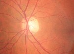 |
| The left eye in this patient demonstrates a thinned and pale neuroretinal rim. |
I made no changes to her therapy at this visit. At her next visit one month later, threshold visual fields demonstrated above and below paracentral scotomata, O.S. greater than O.D., with a significant nasal step O.S. Her IOPs were 15mm Hg in each eye.
Although her glaucoma seemed controlled and stabilized, I asked her to discontinue the Timoptic and begin Travatan (travoprost, Alcon) one drop O.U. h.s.
Discussion
My interns have heard me say numerous times, Dont fix it if it isnt broken. So, why did I choose to fix something that apparently wasnt broken?
Actually, this patients situation was not necessarily broken; it was simply inefficient. Clearly, her glaucomatous optic neuropathy had stabilized, as evidenced by her medical records and her current optic nerve appearance and stable function over many years. She tolerated the medication well and was fully compliant.
In asking the patient about her medication regimen, she reported that she takes all her medications in the morning except for the Timoptic and the Toprol. She said she had always taken the Timoptic h.s., but switched her Toprol to h.s., as it made her tired when taken in the morning. Her GP explained to her why that was the case, and concurred that taking the Toprol at bedtime would result in less daytime fatigue and may even help her sleep better at night, which she found to be true. Both times I saw the patient in the morning. On the second visit, her blood pressure was 112/64, and her pulse rate was 64.
Looking at the patients optic nerve closely, her neuroretinal rims were slightly pale. This may be a long-term effect of pseudophakic pallor, which is certainly plausible. However, perfusion to the optic nerve can also be reduced in glaucoma. Furthermore, a recent study demonstrated that as age increases, blood volume and blood flow to the optic nerve decrease.1
Beta blockers, both systemic (oral) and topical, reduce blood pressure, lower heart rate and decrease cardiac output. She was taking two beta blockers, one topically and one orally, at night. The potential result of this combination is an inordinately low blood pressure (and subsequently inordinately low cardiac output) during sleep, with potentially decreased perfusion to the optic nerve at that same time. A soon-to-be-published study looks at the effects of both topical and oral beta blockers on pulse rates in glaucoma patients.2 The results show that patients not using beta blockers have a higher resting pulse rate than those using topical, oral, or both topical and oral beta blockers, and the resting pulse rates decrease progressively from 76 beats per minute (bpm), to 70 bpm to 65 bpm to 58 bpm, respectively.
Given that this patient is older, uses both systemic and topical beta blockers, and appears clinically to have decreased perfusion to her optic nerves (as manifested by slight optic nerve pallor), I discontinued the topical beta blocker and substituted a once-daily dosage of a prostaglandin. Was this change absolutely necessary? No, but its prudent since it could decrease the likelihood of compromising optic nerve blood flow.
On follow up, the patients IOPs were 12mm Hg O.U., and she is tolerating the Travatan well. She didnt necessarily feel any betterbut I did, even though on the surface it seems that I fixed something that wasnt broken.
1. Boehm AG, Koeller AU, Pillunat LE. The effect of age on optic nerve head blood flow. Invest Ophthal Vis Sci 2005 Apr;46(4):1291-5.
2. Tattersall C, Vernon S, Singh R. Resting pulse rates in a glaucoma clinic: The effect of topical and systemic beta-blocker usage. Eye 2005 Apr 1; [Epub ahead of print].
