 A 55-year-old Hispanic male presented with decreased vision that had persisted for three months. The patient said that his vision has gradually improved since initial onset. He also noted experiencing headaches, but he said that they too have subsided recently.
Prior to this experience, the patient said that he enjoyed good vision with spectacle correction; however, his glasses were no longer helping. Additionally, the patient had been diagnosed with hypertension one month prior to the office visit. Currently, he is taking an oral anti-hypertensive medication and aspirin.
On examination, his best-corrected visual acuity measured 20/40 O.U. Extraocular motility testing was normal. Confrontation fields were full to careful finger counting O.U. His pupils were equally round and reactive to light, with no afferent pupillary defect.
His anterior segment examination was unremarkable O.U. His intraocular pressure measured 13mm Hg O.U. A dilated fundus examination revealed the changes seen in figures 1-4.
|
|
|
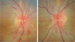
1, 2. Stereo optic nerve photographs of the right and left eye (O.D. left, O.S. right) demonstrate disc edema and nerve fiber layer hemorrhages. |
 |
|
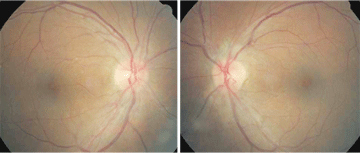
3, 4. Fundus images of our patient (O.D. left, O.S. right). Do you see any retinal changes in addition to papilledema and hemorrhages? |
Take the Retina Quiz
1. What additional testing would be most useful in confirming the diagnosis?
a. Optical coherence tomography (OCT).
b. Color vision testing.
c. Blood pressure measurement.
d. Threshold visual field testing.
2. What is the most likely diagnosis?
a. Non-arteritic anterior optic neuropathy.
b. Malignant hypertension or hypertensive emergency.
c. Idiopathic intracranial hypertension.
d. Neoplasm.
3. How should this patient be managed?
a. Magnetic resonance imaging (MRI).
b. Visual field and neuro-phthalmology consultation.
c. Lumbar puncture and MRI.
d. Immediate referral to the emergency room for blood pressure control.
4. Following treatment, what is the likely visual prognosis for this patient?
a. Very poor.
b. Moderate.
c. Above average.
d. Excellent.
For answers, see below.
Discussion
The fundus photographs show slight blurring of the disc margins O.U. and flame-shaped hemorrhages around both optic nerves. There are retinal folds adjacent to the optic nerve on the temporal side of the left eye, which suggests that the nerve was swollen previously.
Now, it appears that our patient is in the resolution stage. This would explain why his vision has improved during the last three months. It also explains why he no longer experiences headaches. So, what does this mean for our patient?
When taking a further history, our patient reported that he was admitted to the hospital one month before his initial eye exam with significantly elevated blood pressure, severe headache and decreased vision. He reported that his systolic blood pressure (SBP) measured 230mm Hg at hospital admission, but could not remember the diastolic blood pressure (DBP). This information and the associated signs of bilateral disc edema and macular edema suggested that he had experienced a hypertensive emergency with end organ damage to the eye and possible hypertensive encephalopathy.
Considering the available information and observations, the changes seen in our patient are classic for malignant hypertension now called hypertensive emergency. Fortunately, he was already receiving excellent care and on the road to recovery. If, however, this were an initial finding with a history of hypertension and high blood pressure, we would have referred our patient to the emergency room for blood pressure reduction immediately. Still, this situation raises a question regarding blood pressure: How high is too high, and what is an emergency vs. an urgency?
Based on the 1993 classification by the Joint National Committee (JNC) on Prevention, Detection, Evaluation and Treatment of High Blood Pressure, SBP higher than 179mm Hg or DBP higher than 109mm Hg is classified as hypertensive crisis.1 Hypertensive crisis can be further categorized as either hypertensive emergency or hypertensive urgency. End organ damage in the presence of significantly elevated blood pressure is classified as hypertensive emergency. Hypertensive crisis in the absence of end organ damage is classified as hypertensive urgency. End organ damage may manifest in the central nervous system, eye, heart (left ventricular dysfunction) and kidney. Hypertensive encephalopathy is associated with severe headaches, change in mental status, transient convulsions, stupor and coma.2
An estimated 65 million Americans, or more, are hypertensive (blood pressure greater than or equal to 140/90mm Hg).3 Hypertensive crisis can either develop
de novo or arise from underlying primary/secondary hypertension. It is relatively uncommon and occurs in approximately 1% of the hypertensive population.4 The presence and extent of end organ damage is related to the patients average blood pressure, the rate of blood pressure elevation, and the amount of underlying hypertensive damage and arterial/capillary change prior to an acute rise in blood pressure.
However, organ dysfunction is uncommon with a DBP less than 130mm Hg.5 Our patient reported no previous history of hypertension, but it is important to keep in mind that approximately 30% of Americans with hypertension are unaware of their condition.6
During hypertensive crisis, autoregulatory loss of the retinal arterioles leads to a breakdown of the blood retinal barrier, causing plasma and protein leakage. Retinal edema is often accompanied by exudates in a radial pattern, which is referred to as a macular star. Optic nerve head edema occurs from either localized ischemia or elevated intraocular pressure associated with hypertensive encephalopathy that results in loss of axoplasmic flow and subsequent swelling. Retinal hemorrhages are a result of capillary wall necrosis. After adequate control of blood pressure, retinal findings may resolve.7
To circumvent extensive end organ damage and morbidity, patients in hypertensive emergency must have their blood pressure lowered immediately through intravenous infusion of antihypertensive medications.
Conversely, patients with hypertensive urgency should have their blood pressure lowered slowly with oral hypertensive agents over a 24- to 48-hour period to avoid subsequent ischemia and infarction secondary to rapid change in blood pressure and loss of arteriole autoregulation.
At his initial visit, our patient was already being treated and carefully monitored for elevated blood pressure; in our office, his blood pressure measured 125/68mm Hg. We informed our patient of the retinal findings and told him that his vision would improve slowly over the next several months. Then, we informed his primary-care physician of the findings and instructed our patient to return in one month.
At follow-up, the patient reported improved vision, which was best-corrected to 20/25 O.U. Fundus examination revealed resolution of macular edema with retinal pigment epithelium mottling and improved bilateral optic nerve head edema with few surrounding flame hemorrhages O.U. We will continue monitoring the patients vision on a monthly basis until complete resolution of the disc edema. Then, we will monitor the patient regularly over longer intervals.
Thanks to Karina Marcovitch, O.D., resident, and Marissa Perez, O.D., assistant professor at Nova Southeastern University College of Optometry for contributing this case.
1. The fifth report of the Joint National Committee on Detection, Evaluation, and Treatment of High Blood Pressure (JNC V). Arch Intern Med 1993 Jan 25;153(2):154-83.
2. Kitiyakara C. Malignant hypertension and hypertensive emergencies. J Am Soc Nephrol 1998 Jan;9(1):133-42.
3. Fields LE, Burt VL, Cutler JA, et al. The burden of adult hypertension in the
4. Lee AG, Beaver HA. Acute bilateral optic disk edema with a macular star figure in a 12-year-old girl. Surv Ophthalmol Jan-Feb 2002:47(1):42-9.
5. Marik PE, Varon J. Hypertensive crises: challenges and management. Chest 2007 Jun;131(6):1949-62.
6. Hajjar I, Kotchen TA. Trends in prevalence, awareness, treatment, and control of hypertension in the United States, 1988-2000. JAMA 2003 Jul 9;290(2):199-206.
7. Kincaid-Smith P, McMichael J, Murphy EA. The clinical course and pathology of hypertension with papilloedema (malignant hypertension). Q J Med 1958 Jan;27(105):117-53.
Retina Quiz Answers: 1) c; 2) b; 3) d; 4) d
