 A 39-year-old Hispanic female presented for an eye exam before applying for a drivers license. The patient did not think she would pass the required vision test for the license, and decided to have her eyes checked first.
She reported that her left eye had been crossed all her life and that she had very poor vision in that eye. She reported no problems in the right eye. Her medical history was unremarkable.
On examination, her best-corrected visual acuity was 20/20 O.D. and 20/60 O.S. Her wet-refraction was +2.25D O.D. and +4.00D O.S.; this was similar to her current prescription. On motility testing, she had a 30.00 prism diopter esotropia O.S. at distance and near. Confrontation visual fields were full to careful finger counting O.U., and her pupils were equally round and reactive with no afferent pupillary defect. Her anterior segment exam was unremarkable. Intraocular pressure measured 18mm Hg in both eyes.
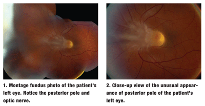
Dilated fundus exam of the right eye was completely normal. In the left eye, however, we observed a fibrous-like band coming off the optic nerve into the vitreous, where it extended nasally to the far peripheral retina (figures 1, 2 and 3). We also performed optical coherence tomography (OCT) on the macula (figures 4 and 5).
 Take the Retina Quiz
1. What does the fibrous band coming off the optic nerve represent?
a. The beginning of a posterior vitreous detachment (PVD).
b. A congenital falciform fold.
c. Failure of the hyaloid artery to regress.
d. Vitreous traction.
2. What does the lesion behind the lens represent?
a. Regressed retinoblastoma.
b. Fibrous hyperplasia.
c. Granuloma.
d. Posterior capsular cataract.
3. What is the likely diagnosis?
a. Persistent fetal vasculature (PFV).
b. Dominant familial exudative vitreoretinopathy (FEVR).
c. Toxocariasis.
d. Retinoblastoma.
4. What is the best treatment option for patients who have this condition?
a. Observation.
b. Vitrectomy and membrane peel.
c. Strabismus surgery.
d. External beam radiation.
5. What form of estropia does this patient likely have?
a. Paralytic estropia.
b. Accommodative estropia.
c. Congenital estropia.
d. Sensory estropia.
For answers, see below.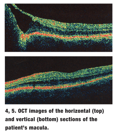 Discussion
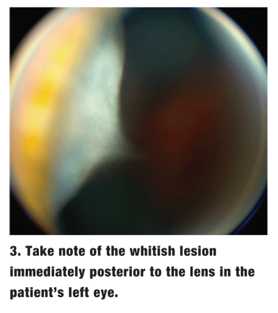
The fibrous-like band that comes off the optic nerve and extends into the vitreous represents the congenital hyaloid artery, which failed to regress. Additionally, vascular remnants are seen within the fibrous band. As noted in the history, the fibrous band extended nasally into the far periphery, where it stopped immediately behind the lens. There, a white, fibrous plaque is seen.
The fibrous plaque is difficult to observe on routine exam and can be overlooked easily. In fact, you almost have to be suspicious that the plaque is present to look for it. To observe it on dilated fundus exam, the patient must look in an extreme gaze to the right while you focus the slit lamp behind the lens.
During indirect ophthalmoscopy, the fibrous plaque can also be seen while looking at the peripheral retina in that location while pulling the condensing lens further away. This allows you to focus more anteriorly and see the plaque.
These findings suggest that the patient has persistent fetal vasculature (PFV), formerly known as persistent hyperplastic primary vitreous (PHPV). PFV results from failure of the primary vitreous and fetal vasculature to regress during normal fetal development. The conditions name was changed to PFV in 1997, as PHPV only sufficently described the more severe form of the disease.1
PFV is more prevalent in infants that have leukocoria in the presence of microphthalmia.1 Upon referral, the infants are usually suspected of retinoblastoma, which is easily ruled out by the presence of both micropthalmia and the persistent hyaloid artery.
In the most severe form of PFV, the fibrous band containing the hyaloid artery can extend from the optic nerve far enough to entirely cover the posterior surface of the lens. This, in turn, causes a vascularized retrolental membrane to form. Tractional retinal detachment involving the posterior pole can also occur.
In the worst cases, patients can develop angle-closure glaucoma, which is the primary reason why vitrectomy is indicted. The clinical spectrum of PFV can vary greatly. The condition presents in two forms: Anterior and posterior.
In the anterior form of PFV, a whitish or pinkish fibrovascular plaque may be present on the posterior surface of the lens. The plaque often appears just nasal to center of the lens. Depending on how large the plaque is, drawn-in ciliary processes can also be seen. The lens is usually clear, but a cataract can develop over time. Persistent papillary membranes and Mittendorf dots are often observed in the anterior form of PFV.
In the posterior form of PFV, a stalk of condensed, or hyperplastic, vitreous attaches at the optic nerve and extends into the vitreous. From there, it can be followed anteriorly to the posterior lens surface or just adjacent to it, as in our patient.
Depending on the severity, there can be considerable traction at the nerve and surrounding retina, possibly leading to retinal dysplasia and retinal folds. Patients may also develop optic nerve hyperplasia. In milder forms of PFV, persistent hyaloid artery remnants and Bergmeister papillae can be seen.
Our patient has the posterior form of PFV. Fortunately, she still has relatively decent visual acuity in the affected eye.
One unanswered question: Just how adversely does this condition affect her visual acuity? An OCT revealed no macular edema; however, there is not a well-developed foveal depression.
Horizontal cut revealed the presence of subretinal fluid and an elevated retina, yet it was difficult to determine how close the measurement came to the center of the fovea. As a result, we do not really know how much the disease affects her visual acuity.
Also, we do not know the relationship between the PFV and her crossed eyes. The initial impression is that the esotropia is a result of sensory deprivation from the PFV. However, she has a moderate amount of hyperopia, which may explain why she has both amblyopia and esotropia. I suspected that the esotropia, reduced visual acuity and retinal changes were all associated with the PFV.
Ultimately, we gave the patient a new prescription for her glasses and filled out the appropriate form for her to get her drivers license. We also referred her for an evaluation for strabismus surgery.
Quiz Answers: 1) c; 2) b; 3) a; 4) c; 5) d.
1. Goldberg MF. Persistent fetal vasculature (PFV): an integrated interpretation of signs and symptoms associated with persistent hyperplastic primary vitreous (PHPV). LIV Edward Jackson Memorial Lecture. Am J Ophthalmol 1997 Nov; 24(5):587-626.
