 As the man in the AT&T commercial might ask, “Which is better: More or less?” Of course, we all like more when it comes to our phone and Internet service. Likewise, we might prefer a bigger office, car or dinner portion at our favorite restaurant. But in some cases, more is definitely not better—especially when it involves the growth or proliferation of our skin into unsightly lesions.
When initially encountering a skin lesion of the eyelid or adnexa, it is crucial to differentiate benign from malignant. Keep in mind that skin cancer remains the most common form of cancer in the United States.1 So, if there is any question or suspicion of malignancy, you must promptly refer the patient for a biopsy evaluation. If the lesion is known or shown to be benign, however, you must then determine its etiology.
|
|
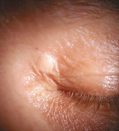
| A 45-year-old woman exhibited xanthelasma of the left upper eyelid. |
Papillomas, xanthelasmas and seborrheic keratoses are benign lesions that often are encountered in optometric practice, and can cause both functional and cosmetic concerns for our patients.
• Papillomas are one of the more common benign epithelial skin lesions, exhibiting a characteristic configuration of lobular projections that can resemble mulberries or cauliflower.2 They may be flat or pedunculated (i.e., on a stalk). Further, the lesions may be solitary or multiple in presentation, and can vary in coloration—although the lesion typically approximates the patient’s skin tone.
Papillomas may be of viral origin. In this instance, they are referred to as verrucae, viral papillomas or warts.3 Those of a non-viral origin are termed acrochordons, squamous papillomas or skin tags.4 Papillomas may occur at any age, although there is a greater predilection toward the viral variety before age 30 and the squamous variety beyond age 30.5
• Xanthelasmas are seen clinically as oval or elongated yellowish plaques located just beneath the skin of the periorbital region. Patients typically are more than 40 years of age, and women are affected nearly twice as often as men.6 The lesions are neither inflammatory nor painful, and there is no tendency toward malignancy; however, the lesions may enlarge and/or coalesce over time. In rare instances, abnormally large xanthelasmas can interfere with lid function.
• Seborrheic keratoses present as sharply demarcated, round- or oval-shaped lesions on the eyelids or adnexa. They usually are elevated, ranging from a few millimeters to several centimeters in diameter. Seborrheic keratoses are classically described as having a “stuck on” quality to them. For this reason, and because they are seen more frequently in elderly patients, they have earned the moniker “barnacles of aging.” The color may vary to extremes, much in the same fashion as papillomas. Nonetheless, most seborrheic keratoses appear a dull brownish-grey. These lesions generally are asymptomatic, but occasionally irritation or trauma may occur with subsequent itching, pain, redness, bleeding or crusting.7
Surgical Treatment Options
The aforementioned benign lesions may be treated in-office in a variety of ways.
• Electrocautery and surgical curettage is an effective treatment option that dermatologists and oculoplastic surgeons often employ for both ocular and non-ocular lesions.8 However, these methods can be painful unless local anesthesia is used, and might yield cosmetically displeasing scarring.
• Cryotherapy with liquid nitrogen is another option—although treating periocular lesions in this fashion presents a significant risk for hypo- or hyperpigmentation (especially in dark-skinned individuals), as well as nerve damage when therapy is too aggressive.8 Also, the inadvertent application of cryogens to the ocular surface can cause severe inflammation and scarring.
• Laser ablation is also an option for many of these lesions, but optometrists are currently restricted from using this class of surgical instrumentation in all but two states (Oklahoma and Kentucky).
Chemical Cautery
Historically, optometrists did not have the ability to manage such lesions via surgical intervention. But today, there is a simple and effective method for removing benign lid and adnexal skin growths: chemical cautery with dichloroacetic acid. This approach is fully within the scope of optometric practice in many states, and is the safest and least invasive way to remove such lesions.
Dichloroacetic acid is a colorless, mildly pungent agent that has both keratolytic and cauterant properties. It can be obtained from a compounding pharmacist at a relatively low price (e.g., $45 from Gale Compounding Pharmacy in Pearson, Texas), or purchased as part of a complete treatment kit, such as the Derma-Cauter-All (approximately $260 from North Pine Enterprises).
The treatment process is extremely straightforward and easy to perform. It may be completed in or out of the slit lamp, but use of magnification generally is advisable. Be sure to wash your hands thoroughly and wear gloves during the procedure.
Prior to treatment, anesthetize the eye(s) with topical 0.5% proparacaine or tetracaine to prevent reflex lacrimation or other irritation from the acid’s fumes. Then, apply a liberal amount of petrolatum around the base of the lesion, extending out at least 2mm to 3mm from the border of the intended treatment area. This helps protect the surrounding skin from incurring collateral damage. Take care not to coat the lesion itself, as this will render the tissue impervious to the acid’s effects.
Next, use the dropper to transfer a small amount of acid from the bottle into a glass vial. Use wooden applicators (which resemble fancy toothpicks) to absorb a small amount of the acid from the glass receptacle, and apply this directly to the lesion. Repeat the process as many times as necessary in order to cover the entire area. Discard used applicators directly into a biohazard bag to avoid unintentional burns or contamination. Advise the patient that the acid usually produces a slight burning or stinging sensation upon application and for a short while thereafter (in practice, this feeling frequently has been compared to an “ant bite”).
When executed correctly, the treated area will turn a milky greyish-white color, indicating that the lesion has been cauterized and the small blood vessels were destroyed. This process causes the tissue to become necrotic and die, leaving behind only normal, viable cells.
In the ensuing days, the patient will notice the lesion becoming darker and scalier, forming an “eschar.” After a week or two, this area will slough off completely to reveal healthy pink tissue that ultimately changes to match the patient’s normal skin tone.
Although not critical, we recommend treating the eschar with a topical antibiotic/steroid ointment (e.g., TobraDex [tobramycin/dexamethasone, Alcon] ung TID to QID) to protect against infection and minimize inflammation and discomfort. Should any portion of the original lesion remain after two weeks, the area may be retreated; however, multiple studies have shown that chemical cautery is effective in eliminating a majority of lesions with just a single application.9,10
While lid “lumps and bumps” may occasionally represent an ominous situation, most of these lesions are benign and merely a cosmetic nuisance for our patients. With proper training in those areas that permit it, optometrists can provide a valuable service for their patients by helping to eliminate these unsightly growths quickly, easily and affordably.
1. US Cancer Statistics Working Group. United States Cancer Statistics: 1999–2010 Incidence and Mortality Web-based Report. CDC and National Cancer Institute; 2013. Available at: www.cdc.gov/uscs. Accessed December 21, 2013.
2. Papilloma. In: Dutton JJ, Gayre GS, Proia AD (eds.). Diagnostic Atlas of Common Eyelid Diseases. New York: CRC Press; 2007:225-6.
3. Verruca vulgaris. In: Dutton JJ, Gayre GS, Proia AD (eds). Diagnostic Atlas of Common Eyelid Diseases. New York: CRC Press; 2007: 258-60.
4. Pariser RJ. Benign neoplasms of the skin. Med Clin North Am. 1998;82(6):1285-307, v-vi.
5. Bellew SG, Quartarolo N, Janniger CK. Childhood warts: an update. Cutis. 2004;73(6):379-84.
6. Gladstone GJ, Myint S. Xanthelasma. In: Fraunfelder FT, Roy FH (eds.). Current Ocular Therapy, 5th Ed. Philadelphia: WB Saunders; 2000: 452-3.
7. Hafner C, Vogt T. Seborrheic keratosis. J Dtsch Dermatol Ges. 2008 Aug;6(8):664-77.
8. Lipke MM. An armamentarium of wart treatments. Clin Med Res. 2006;4(4):273-93.
9. Haygood LJ, Bennett JD, Brodell RT. Treatment of xanthelasma palpebrarum with bichloracetic acid. Dermatol Surg. 1998 Sep;24(9):1027-31.
10. Taner ZM, Taskiran C, Onan AM, et al. Therapeutic value of trichloroacetic acid in the treatment of isolated genital warts on the external female genitalia. J Reprod Med. 2007 Jun;52(6):521-5.
